. 2024 Dec;29(1):2404794.
doi: 10.1080/13510002.2024.2404794. Epub 2024 Sep 23.

WTAP-mediated m6A modification of TRIM22 promotes diabetic nephropathy by inducing mitochondrial dysfunction via ubiquitination of OPA1

Affiliations

WTAP-mediated m6A modification of TRIM22 promotes diabetic nephropathy by inducing mitochondrial dysfunction via ubiquitination of OPA1

Zeng Zhang et al. Redox Rep. 2024 Dec.

Abstract

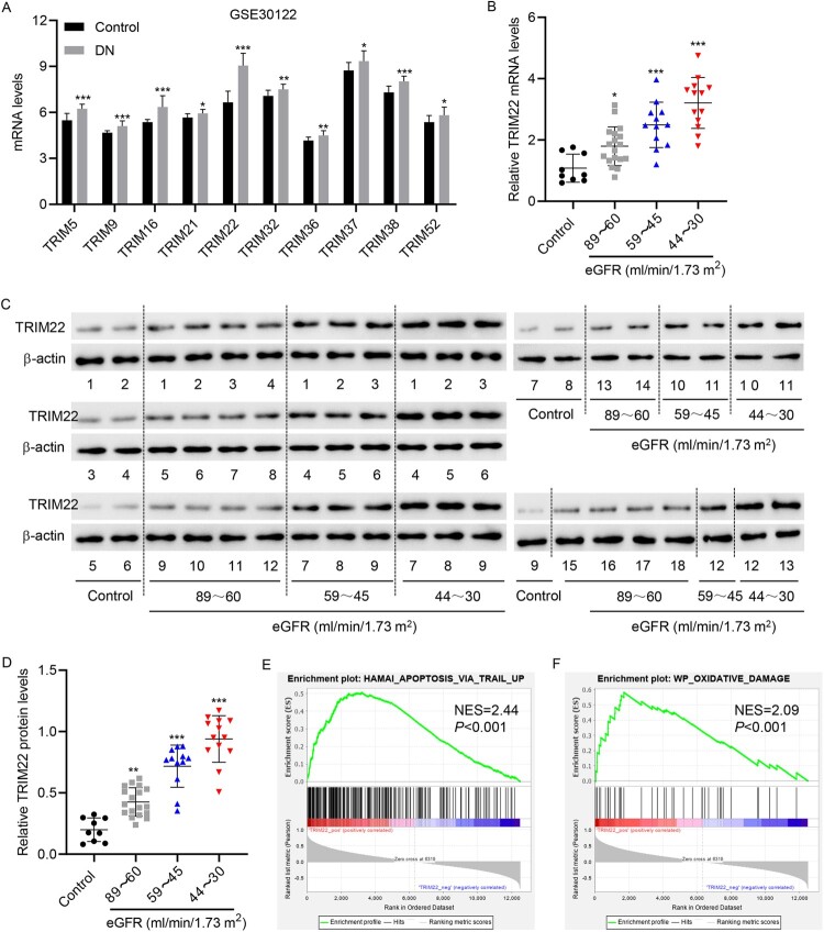
Objectives: Diabetic nephropathy (DN) is one of the most serious microvascular complications of diabetes and is the most common cause of end-stage renal disease. Tripartite motif-containing (TRIM) proteins are a large family of E3 ubiquitin ligases that contribute to protein quality control by regulating the ubiquitin - proteasome system. However, the detailed mechanisms through which various TRIM proteins regulate downstream events have not yet been fully elucidated. The current research aimed to determine the function and mechanism of TRIM22 in DN.

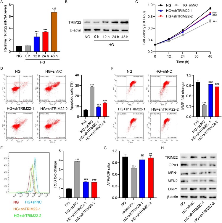
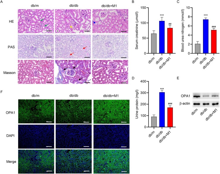
Methods: DN models were established by inducing HK-2 cells using high glucose (HG) and diabetic mice (db/db mice). Cell viability, apoptosis, mitochondrial reactive oxygen species, and mitochondrial membrane potential were detected by Cell Counting Kit-8 and flow cytometry, respectively. Pathological changes were evaluated using hematoxylin and eosin, periodic acid schiff and Masson staining. The binding between TRIM22 and optic atrophy 1 (OPA1) was analyzed using co-immunoprecipitation. The m6A level of TRIM22 5'UTR was detected using RNA immunoprecipitation.

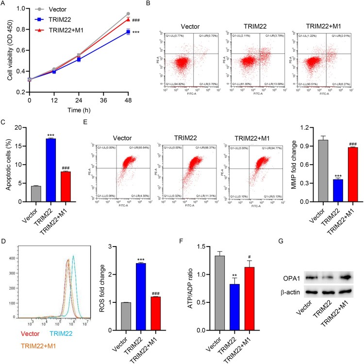
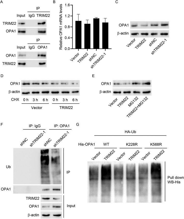
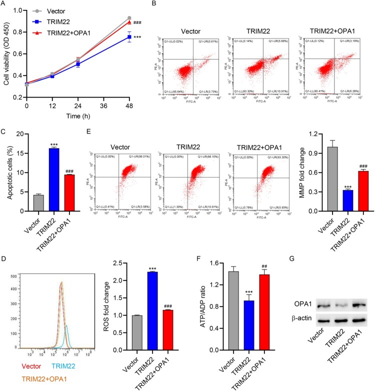
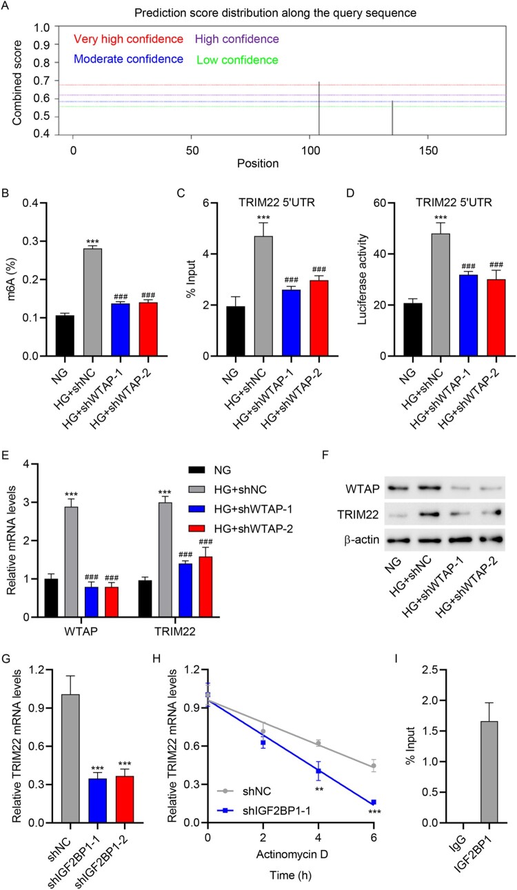
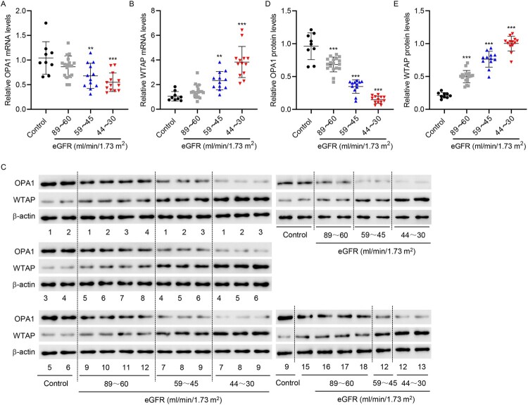
Results: TRIM22 was highly expressed in patients with DN. TRIM22 silencing inhibited HG-induced apoptosis and mitochondrial dysfunction in HK-2 cells. Promoting mitochondrial fusion alleviated TRIM22 overexpression-induced cell apoptosis, mitochondrial dysfunction in HK-2 cells, and kidney damage in mice. Mechanistically, TRIM22 interacted with OPA1 and induced its ubiquitination. Wilms tumor 1-associating protein (WTAP) promoted m6A modification of TRIM22 through the m6A reader insulin-like growth factor 2 mRNA-binding protein 1 (IGF2BP1).

Discussion: TRIM22 silencing inhibited the progression of DN by interacting with OPA1 and inducing its ubiquitination. Furthermore, WTAP promoted m6A modification of TRIM22 via IGF2BP1.

Keywords: OPA1; TRIM22; WTAP; diabetic nephropathy; m6A; mitochondrial dysfunction.


Conflict of interest statement

No potential conflict of interest was reported by the author(s).

Figures

Figure 1.
Figure 1.
TRIM22 expression in patients with DN. (A) The GSE30122 database was used to analyze the expression of TRIM family members in the renal tubules of patients with DN. The renal tubules of patients with DN were collected and divided into three groups according to eGFR (ml/min/1.73 m2) (the 89–60 group: 18 cases; 59–45 group: 12 cases; and 44–30 group: 13 cases). Simultaneously, the renal tubules of nine normal controls undergoing renal puncture were collected. TRIM22 expression was detected using (B) RT-qPCR and (C, D) Western blotting. (E, F) GSEA analysis of the correlation between TRIM22 expression and HAMAI_ APOPTOSIS_ VIA_ TRAIL_ UP and WP_ OXIDATIVE_ DAMAGE signaling pathways. *P < 0.05, **P < 0.01, ***P < 0.001 versus control.
Figure 2.
Figure 2.
TRIM22 silencing inhibited HG-induced apoptosis and mitochondrial dysfunction in HK-2 cells. HK-2 cells were treated with high glucose (HG, 30 mM) (osmotic pressure was controlled with normal glucose concentration 5.5 and 24.5 mM mannitol, NG) to construct a renal tubular injury model of diabetes. TRIM22 expression was detected using (A) RT-qPCR and (B) Western blotting at 0, 6, 12, 24, and 48 h. HK-2 cells were transduced with TRIM22 shRNA lentivirus (shTRIM22-1 and shTRIM22-2) or scramble shRNA (shNC) and stimulated with HG for 48 h. (C) CCK-8 was used to determine cell viability. (D) Flow cytometry was used to detect cell apoptosis, (E) mitochondrial ROS, and (F) MMP. (G) Biochemical assay was used to determine the ATP/ADP ratio. (H) Western blotting was used to detect TRIM22, OPA1, MFN1, MFN2, and DRP1 expression. ***P < 0.001 versus NG; ##P < 0.01, ###P < 0.001 versus HG + shNC.
Figure 3.
Figure 3.
Promotion of mitochondrial fusion relieved TRIM22 overexpression-induced apoptosis and mitochondrial dysfunction in HK-2 cells. HK-2 cells transduced with TRIM22 expression vector or blank vector were treated with 10 μM mitochondrial fusion inducer M1 alone or in combination for 48 h. (A) CCK-8 was used to determine cell viability. (B, C) Flow cytometry was used to detect cell apoptosis, (D) mitochondrial ROS, and (E) MMP. (F) Biochemical assay was used to determine the ATP/ADP ratio. (G) Western blotting was used to determine OPA1 expression. ***P < 0.001 versus vector; #P < 0.05, ###P < 0.001 versus TRIM22.
Figure 4.
Figure 4.
Promotion of mitochondrial fusion reversed kidney injury in mice. Eight-week-old diabetic mice were divided into a control group (db/m), model group (db/db), and model + mitochondrion fusion inducer M1 (db/db + M1) group that received intervention for 4 weeks. (A) HE, PAS, and Masson staining were used to analyze the pathological changes in renal tissues (scale bar, 50 μm). HE staining identified normal proximal tubules with narrow and irregular lumena, unclear cell boundaries, and the presence of brush border structures (green arrow) and abnormal proximal tubules with tubular dilatation (*), atrophy (yellow arrow), and loss of brush border integrity (blue arrow). PAS staining was used to identified renal tubular dilatation (red arrow). Masson staining identified extracellular matrix deposition (black arrow). Biochemical detection of (B) creatinine, (C) urea nitrogen and (D) urinary protein. (E) Western blotting and (F) immunofluorescence staining were used to determine OPA1 expression in renal tissues (scale bar, 100 μm). ***P < 0.001 versus db/m; ##P < 0.01, ###P < 0.001 versus db/db.
Figure 5.
Figure 5.
TRIM22 interacted with OPA1 and induced its ubiquitination. (A) Co-IP was used to detect the binding activity of TRIM22 and OPA1. TRIM22 shRNA lentivirus (shTRIM22-1), scramble shRNA (shNC), TRIM22 expression vector or blank vector was transfected into 293 T cells. (B) RT-qPCR and (C) Western blotting were used to detect OPA1 expression. (D) 293 T cells transfected with TRIM22 expression vector or blank vector were treated with protein synthesis inhibitor CHX, and Western blotting was used to determine OPA1 expression. (E) TRIM22 expression vector or blank vector were transfected into 293 T cells, which were subsequently treated with MG132 alone or in combination for 4 h and analyzed for OPA1 expression using Western blotting. (F) TRIM22 shRNA lentivirus (shTRIM22-1) or scramble shRNA (shNC) were transfected into 293 T cells, after which IP and Western blotting were used to detect the ubiquitination. (G) Cells were co-transfected with the His-OPA1 (WT) or mutant His-OPA1 constructs (K228R and K568R) along with TRIM22 expression vector or blank vector and HA-Ub construct and then analyzed using the pull-down assay.
Figure 6.
Figure 6.
OPA1 overexpression relieved TRIM22 overexpression-induced apoptosis and mitochondrial dysfunction in HK-2 cells. Co-transduction of TRIM22 and OPA1 expression vector into HK-2 cells for 48 h. (A) CCK-8 was used to determine cell viability. (B, C) Flow cytometry was used to detect cell apoptosis, (D) mitochondrial ROS, and (E) MMP. (F) Biochemical assay was used to determine the ATP/ADP ratio. (G) Western blotting was used to detect OPA1 expression. ***P < 0.001 versus vector; ###P < 0.001 versus TRIM22.
Figure 7.
Figure 7.
WTAP promoted the m6A modification of TRIM22 via the m6A reader IGF2BP1. (A) The SRAMP website was used to predict the m6A modification of TRIM22. HK-2 cells were transduced with WTAP shRNA lentivirus (shWTAP-1 and shWTAP-2) or scramble shRNA (shNC) and stimulated with HG for 48 h. (B) ELISA was used to determine m6A levels. (C) RIP was used to determine TRIM22 mRNA 5′UTR m6A levels. (D) Luciferase reporter gene assay was used to determine TRIM22 mRNA 5′UTR activity. (E) RT-qPCR and (F) Western blotting were used to determine the expression of WTAP and TRIM22. (G) HK-2 cells were transduced with IGF2BP1 shRNA lentivirus (shIGF2BP1-1 and shIGF2BP1-2) or scramble shRNA (shNC) and analyzed for TRIM22 expression using RT-qPCR. (H) HK-2 cells were transduced with IGF2BP1 shRNA lentivirus (shIGF2BP1-1) or scramble shRNA (shNC), followed by actinomycin D treatment for 0, 2, 4, and 6 h. The transcription level of TRIM22 was determined using RT-qPCR. (I) RIP-PCR was used to detect the binding of IGF2BP1 to TRIM22 mRNA 5′UTR. **P < 0.01, ***P < 0.001 versus NG or shNC; ###P < 0.001 versus HG + shNC.
Figure 8.
Figure 8.
OPA1 and WTAP expression in patients with DN. The renal tubules of DN patients were collected and divided into three groups according to eGFR (ml/min/1.73 m2) (The 89–60 group: 18 cases; 59–45 group: 12 cases; and 44–30 group: 13 cases). Simultaneously, the renal tubules of nine normal controls undergoing renal puncture were collected. (A, B) RT-qPCR and (C – E) Western blotting were used to determine OPA1 and WTAP expression. **P < 0.01, ***P < 0.001 versus control.

References

    1. Pan S, Li Z, Wang Y, et al. A Comprehensive weighted gene co-expression network analysis uncovers potential targets in diabetic kidney disease. J Transl Int Med. 2023;10:359–368. doi: 10.2478/jtim-2022-0053 - DOI - PMC - PubMed
    1. Alicic RZ, Rooney MT, Tuttle KR.. Diabetic kidney disease: challenges, progress, and possibilities. Clin J Am Soc Nephrol. 2017;12:2032–2045. doi: 10.2215/CJN.11491116 - DOI - PMC - PubMed
    1. Li J, Shu L, Jiang Q, et al. Oridonin ameliorates renal fibrosis in diabetic nephropathy by inhibiting the Wnt/β-catenin signaling pathway. Ren Fail. 2024;46(1):2347462. doi: 10.1080/0886022X.2024.2347462 - DOI - PMC - PubMed
    1. Amorim JA, Coppotelli G, Rolo AP, et al. Mitochondrial and metabolic dysfunction in ageing and age-related diseases. Nat Rev Endocrinol. 2022;18:243–58. doi: 10.1038/s41574-021-00626-7 - DOI - PMC - PubMed
    1. Brand MD, Nicholls DG.. Assessing mitochondrial dysfunction in cells. Biochem J. 2011;435:297–312. doi: 10.1042/BJ20110162 - DOI - PMC - PubMed

MeSH terms