. 2024 Dec 16;25(24):e202400631.
doi: 10.1002/cbic.202400631. Epub 2024 Nov 9.

Cavity-Based Discovery of New Fatty Acid Photodecarboxylases

Affiliations

Cavity-Based Discovery of New Fatty Acid Photodecarboxylases

Stefan Simić et al. Chembiochem. .

Abstract

Light-dependent fatty acid photodecarboxylases (FAPs) hold significant potential for biotechnology, due to their capability to produce alka(e)nes directly from the corresponding (un)saturated natural fatty acids requiring light as the only reagent. This study expands the family of FAPs through cavity-based enzyme discovery methods. Thirty enzyme candidates with potential photodecarboxylation activity were identified by matching the cavities of four related template structures against the Protein Data Bank's flavoproteins, a library of proteins identified via the Foldseek Search Server, and homology models of sequences resulting from BLAST. Subsequent docking experiments narrowed this library to ten promising enzymes, which were expressed and assessed in vitro, identifying four photodecarboxylases. Out of these enzymes, the GMC oxidoreductase from Coccomyxa sp. Obi (CoFAP) was characterized in detail, which revealed high activity in the decarboxylation reactions of palmitic acid and octanoic acid and a broad pH tolerance (pH 6.5-9.5).

Keywords: Biocatalysis; Enzyme discovery; Photocatalysis; Photodecarboxylase.


Conflict of interest statement

The authors declare no conflict of interest.

Figures

Figure 1
Figure 1
A: Photodecarboxylation or fatty acids catalyzed by CvFAP. B: Workflow for the cavity‐based discovery of new photodecarboxylases consisting of the procreation of four template point‐clouds (exemplary point clouds are shown), matching experiments with three sources of protein structures (the FoldSeek structures overlap with the PDB structures), high throughput docking experiments and in vitro testing.
Figure 2
Figure 2
A: Active site of CvFAP (PDB ID: 6YRU) with bound stearic acid (dark grey) and the truncated substrate that was used for cavity procreation (blue); B: Point cloud in the active site of CvFAP (PDB ID: 6YRU); C: False‐positive match: this point cloud is not aligned with the FAD (PDB ID: 2GVC); D: False‐positive match: the point clouds of CvFAP (PDB ID: 6YRU) and the target structure (PDB ID: 5FNO) are aligned, but the flavins point into different directions (i. e., the point clouds match, but the cofactor is on the opposite side of the point cloud). E: Alignment of CvFAP (PDB ID: 6YS1) and COFAP (BLAST candidate homology model). F: Alignment of CvFAP (PDB: 6YRU) and the Foldseek hit (PDB ID: 4HA6). The structure of CvFAP (blue) has a similar orientation in all figures. Any aligned structure is depicted in light grey.
Figure 3
Figure 3
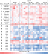
The combined scores (|E/d|) in the docking between candidate cavities and set of carboxylic acid ligands shown as heat map. For a list of the sum of the combined scores, see Table S3 [a] Entries 1–12 show GenBank Protein IDs, entry 13 shows a UniProt ID and entries 14–30 show PDB IDs. [b] The combined scores were calculated as an absolute value of the ratio of energy (kcal⋅mol−1) and distance (COOH−N5, Å). For each ligand, the cluster with minimal distance and the binding energy corresponding to that cluster were taken. [c] oxo, alc: ketones or alcohols; amine+alc.: 1,2‐hydoxyamines. KAF5834678.1: DsFAP; KAG2498954.1: EdFAP; XP_002948047.1: VcFAP; BDA51473.1: COFAP; KAI8103262.1: PiFAP.
Scheme 1
Scheme 1
Substrates investigated for photodecarboxylation activity of enzyme candidates. Enzyme candidates showing activity are enlisted below the substrate structures. In contrast to the dockings which were done with the individual enantiomers of the ligands (see Figure S6) the substrates were used as racemic mixtures, as depicted in the biotransformations. Substrate all‐rac22 corresponds to the ligands 22–25; substrate rac18 corresponds to ligands 18 and 19; and substrate rac16 corresponds to ligands 16 and 17. [a] Reaction conditions: Substrate (10 mM), DMSO (10 % v/v), KPi buffer (100 mM, pH 8, 900 μL total volume) and lyophilized cell lysate (10 mg mL−1, for individual enzyme amounts, see Table S5) were illuminated at 455 nm (36 μmol L−1 s−1), 25 °C, 500 rpm, for 20 h. [b] Reaction conditions: Substrate (10 mM), KPi buffer (100 mM, pH 8, 1 mL total volume) and lyophilized cell lysate (10 mg mL−1) were illuminated under identical conditions as shown above. The buffer was purged with N2 for 1 h and the reactions were performed under N2 atmosphere. All samples were prepared in duplicates.
Figure 4
Figure 4
pH profile of fatty‐acid photodecarboxylase from Coccomyxa sp. Obi (COFAP) displayed as conversion in the photodecarboxylation of palmitic acid. Reaction conditions: palmitic acid (10 mM), DMSO (10 % v/v), buffer (900 μL), COFAP (10 mg mL−1 lyophilized cell lysate, containing 0.1 mg mL−1 COFAP, estimated by SDS PAGE densitometry), total volume 1 mL. Illumination was performed at 455 nm (36 μmol L−1 s−1), 25 °C, 500 rpm, for 20 h. All data points represent mean values of duplicate determinations.
Figure 5
Figure 5
Photodecarboxylation of fatty acids catalyzed by fatty‐acid photodecarboxylase from Coccomyxa sp. Obi (COFAP). Reaction conditions: fatty acid (10 mM), DMSO (10 % v/v), Tris⋅HCl buffer (100 mM, pH 8.5, 900 μL), COFAP (10 mg mL−1 lyophilized cell lysate, containing 0.22 mg mL−1 COFAP, estimated by SDS PAGE densitometry), total volume 1 mL. Illumination was performed at 455 nm (36 μmol L−1 s−1), 25 °C, 500 rpm, for 20 h. For C18 : 1 only substrate depletion was measured. All data points represent mean values of duplicate or triplicate determinations.

Similar articles

References

    1. Sorigue D., Legeret B., Cuine S., Blangy S., Moulin S., Billon E., Richaud P., Brugiere S., Coute Y., Nurizzo D., Muller P., Brettel K., Pignol D., Arnoux P., Li-Beisson Y., Peltier G., Beisson F., Science 2017, 357, 903–907. - PubMed
    1. Schmermund L., Jurkas V., Ozgen F. F., Barone G. D., Büchsenschütz H. C., Winkler C. K., Schmidt S., Kourist R., Kroutil W., ACS Catal. 2019, 9, 4115–4144.
    1. Alphand V., van Berkel W. J. H., Jurkas V., Kara S., Kourist R., Kroutil W., Mascia F., Nowaczyk M. M., Paul C. E., Schmidt S., Spasic J., Tamagnini P., Winkler C. K., ChemPhotoChem 2023, 7, e202200325.
    1. Emmanuel M. A., Bender S. G., Bilodeau C., Carceller J. M., DeHovitz J. S., Fu H., Liu Y., Nicholls B. T., Ouyang Y., Page C. G., Qiao T., Raps F. C., Sorigue D. R., Sun S. Z., Turek-Herman J., Ye Y., Rivas-Souchet A., Cao J., Hyster T. K., Chem. Rev. 2023, 123, 5459–5520. - PMC - PubMed
    1. Taylor A., Heyes D. J., Scrutton N. S., Curr. Opin. Struct. Biol. 2022, 77, 102491. - PubMed

LinkOut - more resources