Case Reports
. 2024 Sep 9:11:1372831.
doi: 10.3389/fmed.2024.1372831. eCollection 2024.

Case reports: Intraoperative migratory retinal venous thrombus in proliferative diabetic retinopathy

Affiliations
Case Reports

Case reports: Intraoperative migratory retinal venous thrombus in proliferative diabetic retinopathy

Danni Lyu et al. Front Med (Lausanne). .

Abstract

Purpose: This study aimed to study the characteristics, possible causes, and clinical implications of intraoperative migratory retinal venous thrombus in proliferative diabetic retinopathy (PDR).

Cases: Two middle-aged Chinese patients with diabetes mellitus presented with blurred vision and were diagnosed with PDR and tractional retinal detachment (TRD). An interesting phenomenon was observed during pars plana vitrectomy in both patients. Movement of tiny white thrombi and interruption of blood flow were observed in a branch of the central retinal vein when the vein was pulled at the time of fibrovascular membrane delamination and disappeared with the elimination of retinal traction after finishing the process of delamination. Laboratory studies revealed abnormal erythrocyte sedimentation rate, fibrinogen, D-dimer, international normalized ratio, and IgA anti-β2-glycoprotein I in one patient and elevated fibrinogen and IgA anticardiolipin in the other. Follow-up examinations at 1 week, 1, 3, and 6 months postoperatively showed good prognosis. Fluorescein fundus angiography at 1 month postoperatively showed neither embolus sign nor prolonged venous filling time in both patients.

Discussion: Local blood stasis of the retinal vein persistently dragged by the fibrovascular membrane may result in thrombogenesis, and traction of the retina during the delamination process may lead to the movement of thrombi. On the other hand, endothelial injury and disordered local blood stasis during delamination may also activate the biological coagulation process and instant thrombus formation. As well, antiphospholipid antibodies may also be a risk factor of ocular thrombogenesis.

Conclusion: This study provides the first videos recording migratory thrombus in terminal vessels, which indicates that fibrovascular membrane in PDR can lead to thrombogenesis due to dragging and hemostasis of the involved retinal vein. PDR patients with fibrovascular membranes may benefit from early relief of vascular traction through fibrovascular membrane delamination.

Keywords: fibrovascular membrane delamination; migratory retinal venous thrombus; proliferative diabetic retinopathy; tractional retinal detachment; vitrectomy.


Conflict of interest statement

The authors declare that the research was conducted in the absence of any commercial or financial relationships that could be construed as a potential conflict of interest.

Figures

Figure 1
Figure 1
Preoperative examination of Patient 1. (A) Ultra-widefield fundus photography of the right eye revealed vast preretinal hemorrhage in the posterior polar and superior regions, together with scattered intraretinal hemorrhage spots and asteroid hyalosis in the periphery. (B) Ultra-widefield fundus photography of the left eye revealed microaneurysms and retinal hemorrhage in all quadrants of the fundus as well as a few hard exudates in the posterior polar retina. (C) Ultrasound of the right eye revealed vitreous opacity and tractional retinal detachment.
Figure 2
Figure 2
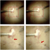
Intraoperative observation of migratory retinal venous thrombus in Patient 1. Rapid movement of tiny white thrombi was observed in the superotemporal branch of the central retinal vein at the time of fibrovascular membrane delamination. The upper two images displayed the full view, while the lower two images displayed the magnified view. Red arrows indicated the migratory retinal venous thrombus.
Figure 3
Figure 3
Preoperative examination of Patient 2. (A) Ultra-widefield fundus photography of the right eye revealed evident tractional retinal detachment with the involvement of the macular area, together with previous PRP spots in the periphery. (B) Ultra-widefield fundus photography of the left eye revealed previous PRP spots in the periphery. (C) Ultrasound of the right eye revealed evident tractional retinal detachment and slight hemorrhagic vitreous opacity. (D) OCT of the right eye revealed retinal detachment and epiretinal membrane in the macular area.
Figure 4
Figure 4
Intraoperative observation of migratory retinal venous thrombus and blood flow interruption in Patient 2. (A) White migratory thrombi with a similar diameter to the vessel were observed during fibrovascular membrane delamination. (B) Interruption of blood flow was observed with evident segmental whitening of the vein, which appeared during the process of membrane delamination. The upper images displayed the full view, while the lower images displayed the magnified view. Red arrows indicate the migratory retinal venous thrombus. Red arrowheads indicate blood flow interruption.
Figure 5
Figure 5
Postoperative examination of Patient 1 at 3 months. Anterior segment photography (A) revealed no positive findings. Ultra-widefield fundus photography (B) and ultrasound (C) revealed a well-attached retina.
Figure 6
Figure 6
Postoperative examination of Patient 2 at 6 months. Anterior segment photography (A) revealed no positive findings. Slight local subretinal fluid was discovered by OCT (B), yet no obvious retinal detachment was detected according to ultra-widefield fundus photography (C) and ultrasound (D).
Figure 7
Figure 7
FFA of Patients 1 and 2 at 1 month after surgery. (A) Venous phase (at 20 s) of FFA showed normal venous filling and no embolus sign in Patient 1. (B) Arteriovenous phase (at 22 s) of FFA showed delayed venous filling without embolus sign in Patient 2. Red arrow indicates the location of the superotemporal branch of the central retinal vein.

References

    1. Chen Z, Chen B, Hu P, Liu H, Zheng D. A preliminary observation on rod cell photobiomodulation in treating diabetic macular edema. Adv Ophthalmol Pract Res. (2022) 2:100051. doi: 10.1016/j.aopr.2022.100051, PMID: - DOI - PMC - PubMed
    1. Antonetti DA, Klein R, Gardner TW. Diabetic retinopathy. N Engl J Med. (2012) 366:1227–39. doi: 10.1056/NEJMra1005073 - DOI - PubMed
    1. Aiello LP, Avery RL, Arrigg PG, Keyt BA, Jampel HD, Shah ST, et al. . Vascular endothelial growth factor in ocular fluid of patients with diabetic retinopathy and other retinal disorders. N Engl J Med. (1994) 331:1480–7. doi: 10.1056/NEJM199412013312203, PMID: - DOI - PubMed
    1. Alkarithi G, Duval C, Shi Y, Macrae FL, Ariens RAS. Thrombus structural composition in cardiovascular disease. Arterioscler Thromb Vasc Biol. (2021) 41:2370–83. doi: 10.1161/ATVBAHA.120.315754, PMID: - DOI - PMC - PubMed
    1. Boeri D, Maiello M, Lorenzi M. Increased prevalence of microthromboses in retinal capillaries of diabetic individuals. Diabetes. (2001) 50:1432–9. doi: 10.2337/diabetes.50.6.1432, PMID: - DOI - PubMed

Publication types

LinkOut - more resources