[Preprint]. 2024 Sep 13:2024.09.12.610282.
doi: 10.1101/2024.09.12.610282.

Impact of anatomic variability and other vascular factors on lamina cribrosa hypoxia

Affiliations

Impact of anatomic variability and other vascular factors on lamina cribrosa hypoxia

Yuankai Lu et al. bioRxiv. .

Abstract

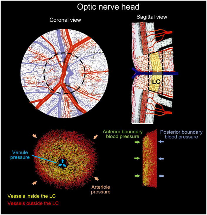
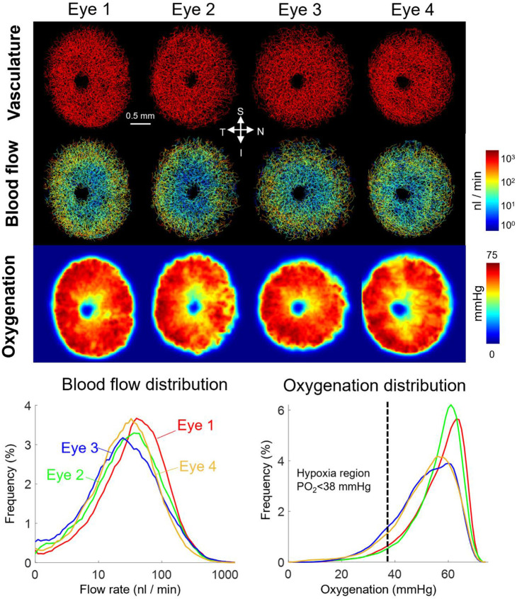
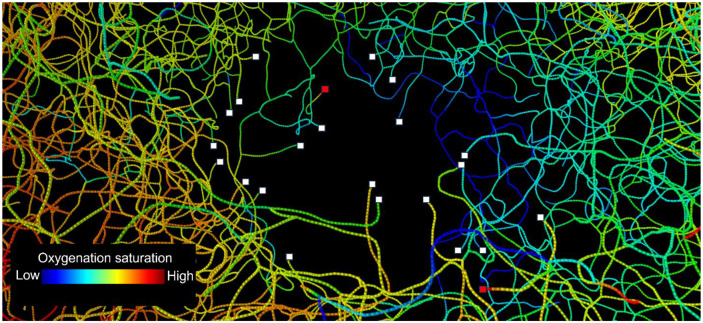
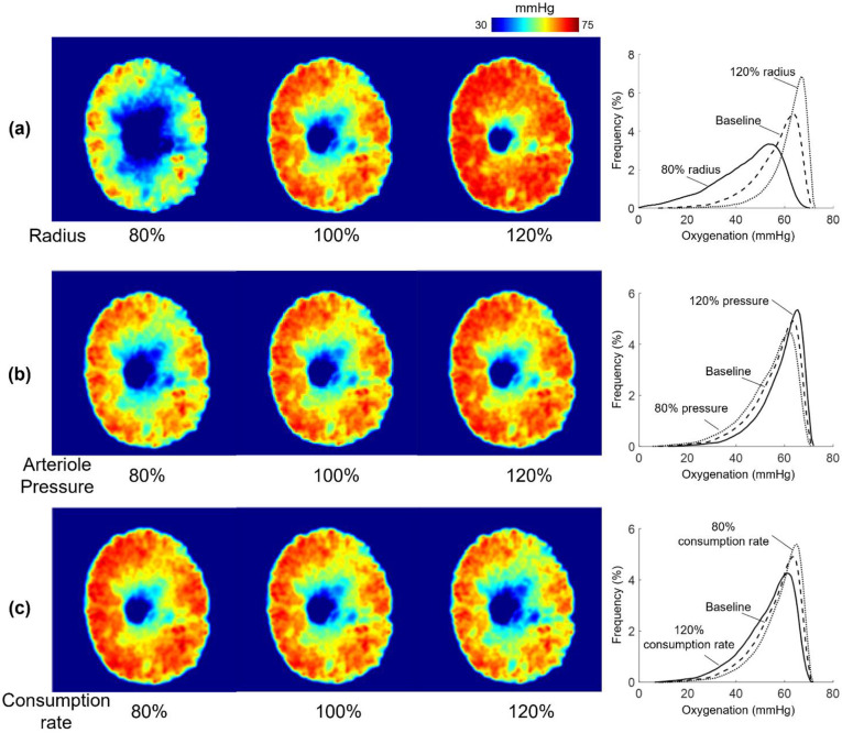
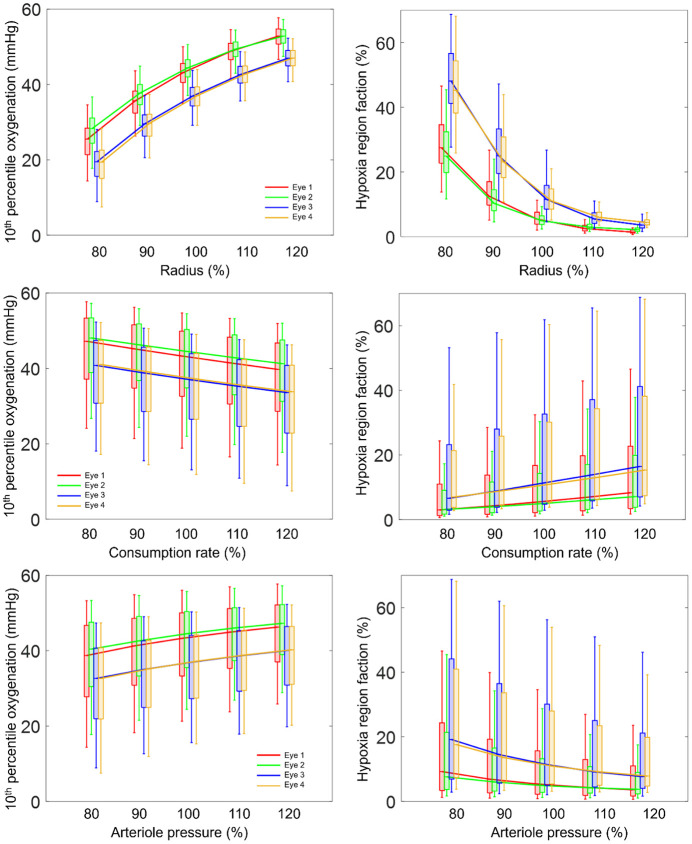
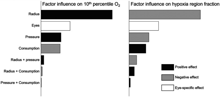
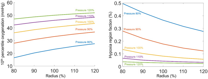
Insufficient oxygenation in the lamina cribrosa (LC) may contribute to axonal damage and glaucomatous vision loss. To understand the range of susceptibilities to glaucoma, we aimed to identify key factors influencing LC oxygenation and examine if these factors vary with anatomical differences between eyes. We reconstructed 3D, eye-specific LC vessel networks from histological sections of four healthy monkey eyes. For each network, we generated 125 models varying vessel radius, oxygen consumption rate, and arteriole perfusion pressure. Using hemodynamic and oxygen supply modeling, we predicted blood flow distribution and tissue oxygenation in the LC. ANOVA assessed the significance of each parameter. Our results showed that vessel radius had the greatest influence on LC oxygenation, followed by anatomical variations. Arteriole perfusion pressure and oxygen consumption rate were the third and fourth most influential factors, respectively. The LC regions are well perfused under baseline conditions. These findings highlight the importance of vessel radius and anatomical variation in LC oxygenation, providing insights into LC physiology and pathology. Pathologies affecting vessel radius may increase the risk of LC hypoxia, and anatomical variations could influence susceptibility. Conversely, increased oxygen consumption rates had minimal effects, suggesting that higher metabolic demands, such as those needed to maintain intracellular transport despite elevated intraocular pressure, have limited impact on LC oxygenation.

Keywords: blood flow; glaucoma; hemodynamics; lamina cribrosa; oxygenation; vasculature.


Conflict of interest statement

Conflict of Interest: none

Figures

Figure 1.
Figure 1.
(Top) Diagram of the ONH adapted from [64]. Our model represents the vessels within the scleral canal, included the whole LC region, and some of the pre-laminar and retro-laminar regions. Black dashed lines represent the model boundaries, yellow area represents the LC region. (Bottom) An example eye-specific vessel network. To improve flow boundary conditions the region reconstructed extended beyond the LC. Vessels within the LC are shown colored in yellow. Vessels reconstructed but outside the LC are shown in red. The network is labeled to illustrate the blood pressure boundary condition settings. Four blood pressure conditions were assigned at the peripheral, central, anterior, and posterior boundaries of the model. See the main text for the rationale and details on how these pressures were assigned.
Figure 2.
Figure 2.
Vascular geometry, baseline hemodynamics, and baseline oxygenation of four eyes. Top: Vascular geometry and maps of blood flow and oxygenation. Bottom: Distributions of blood flow and oxygenation in the LC region. Blood flow and oxygenation exhibited similar features and tendencies across all eyes. Higher flow rates and oxygenation occurred at the periphery of the LC region, and lower flow rates and oxygenation were observed at the center. Interestingly, the distribution curves of blood flow and oxygenation showed different patterns. Blood flow exhibited significant variation across the entire LC, ranging from 1 to 1000 nl/min, while the LC was consistently well-provided with oxygen. Oxygen tensions remain consistently high throughout most of the LC region, with minimal regions experiencing hypoxic conditions.
Figure 3.
Figure 3.
A screenshot of the video illustrating red blood cell transport in the blood vessels. The region shown is a close-up of the drainage at the central retinal vessels. Colors represent the blood oxygen saturation. The dots along each vessel represent the red blood cells. White and red squares represent blood flow outlets and inlets, respectively. The vessel ends in the center of the LC serve as flow outlets, according to the boundary conditions of flow drainage through the center retinal vein. Vessel ends that are not draining are because they end at the model anterior/posterior boundary (see Figure 1) and thus flow can be to/from the LC. Notably, the blood oxygen saturation exhibits an asymmetric pattern at the center, with lower oxygenation observed on the right side (Nasal side).
Figure 4.
Figure 4.
Oxygenation distribution across various radii, consumption rates, and arteriole pressures for eye 1. (a) Oxygenation at different radii. (b) Oxygenation at different Arteriole pressures. (c) Oxygenation at different consumption rates. Three levels—low (80%), baseline (100%), and high (120%)—were selected to illustrate the impact of each parameter. Radius demonstrated the strongest positive influence on oxygenation, while consumption rate and arteriole pressure had minor effects. Most hypoxia regions were observed near the center of the LC across all parametric scenarios.
Figure 5.
Figure 5.
Boxplots showing the factor influences on the 10th percentile oxygenation and hypoxia region fraction in the lamina cribrosa across all eyes. The top and bottom edges of each box are the upper and lower quartiles (25th and 75th percentiles), while the line inside of each box is the sample median (50th percentiles), respectively. The end of the whiskers shows the minimum and maximum. To aid visualization we added lines connecting the median values. Vessel radius shows the strongest positive relationship with the 10th percentile oxygenation and the strongest negative relationship with the hypoxia region fraction. The variation observed among different eyes exceeds the oxygenation difference between scenarios with 80% consumption rate/arteriole pressure and those with 120% consumption rate/arteriole pressure. Interestingly, Eyes 1 and 2 exhibited similar LC oxygenation across all parametric cases, while Eyes 3 and 4 also showed similar patterns to each other but differed from Eyes 1 and 2. The changes in LC oxygenation in response to the parameters, or in other words, the sensitivity to parameters, were consistent across all eyes.
Figure 6.
Figure 6.
Bar chart showing the ranking of factors and interactions according to their influence on the 10th percentile oxygenation and hypoxia region fraction in the LC, as determined by ANOVA. The vessel radius was strongest influence factor, followed by the eye, arteriole pressure, and consumption rate. Interactions between the parameters show minor contributions to the LC oxygenation.
Figure 7.
Figure 7.
Interactions between vessel radius and arteriole pressure on LC oxygenation. Regarding the 10th percentile oxygenation, the curves for different pressures were nearly parallel, suggesting a weak interaction between the radius and pressure parameters. For hypoxia region fraction, the influence of the pressure was stronger when the radius was low.

Similar articles

References

    1. Quigley H., and Anderson D. R., 1976, "The dynamics and location of axonal transport blockade by acute intraocular pressure elevation in primate optic nerve," Investigative ophthalmology & visual science, 15(8), pp. 606–616. - PubMed
    1. Quigley H. A., Nickells R. W., Kerrigan L. A., Pease M. E., Thibault D. J., and Zack D. J., 1995, "Retinal ganglion cell death in experimental glaucoma and after axotomy occurs by apoptosis," Investigative ophthalmology & visual science, 36(5), pp. 774–786. - PubMed
    1. Howell G. R., Libby R. T., Jakobs T. C., Smith R. S., Phalan F. C., Barter J. W., Barbay J. M., Marchant J. K., Mahesh N., and Porciatti V., 2007, "Axons of retinal ganglion cells are insulted in the optic nerve early in DBA/2J glaucoma," The Journal of cell biology, 179(7), pp. 1523–1537. - PMC - PubMed
    1. Quigley H., 2011, "vol. 377," Glaucoma Lancet, pp. 1367–1377. - PubMed
    1. Brazile B. L., Yang B., Waxman S., Lam P., Voorhees A. P., Hua Y., Loewen R. T., Loewen N. A., Rizzo J. F., and Jakobs T., 2020, "Lamina cribrosa capillaries straighten as intraocular pressure increases," Investigative Ophthalmology & Visual Science, 61(12), pp. 2–2. - PMC - PubMed

Publication types

LinkOut - more resources