[Preprint]. 2024 Sep 15:2024.09.12.612721.
doi: 10.1101/2024.09.12.612721.

"Mitotic" kinesin-5 is a dynamic brake for axonal growth

Affiliations

"Mitotic" kinesin-5 is a dynamic brake for axonal growth

Wen Lu et al. bioRxiv. .

Update in

Abstract

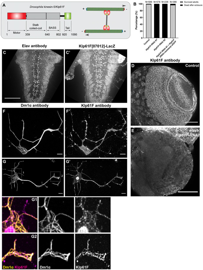
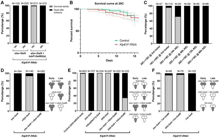
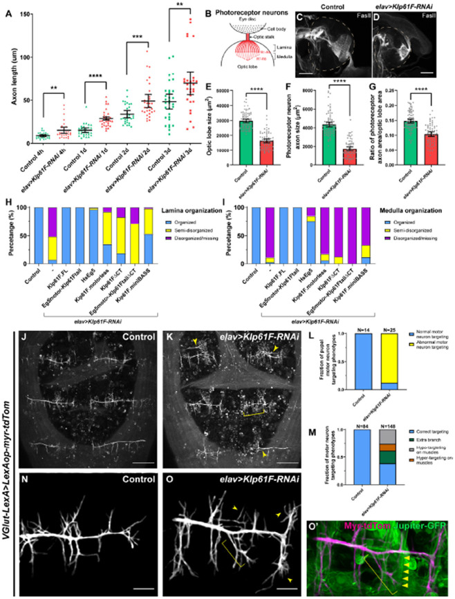
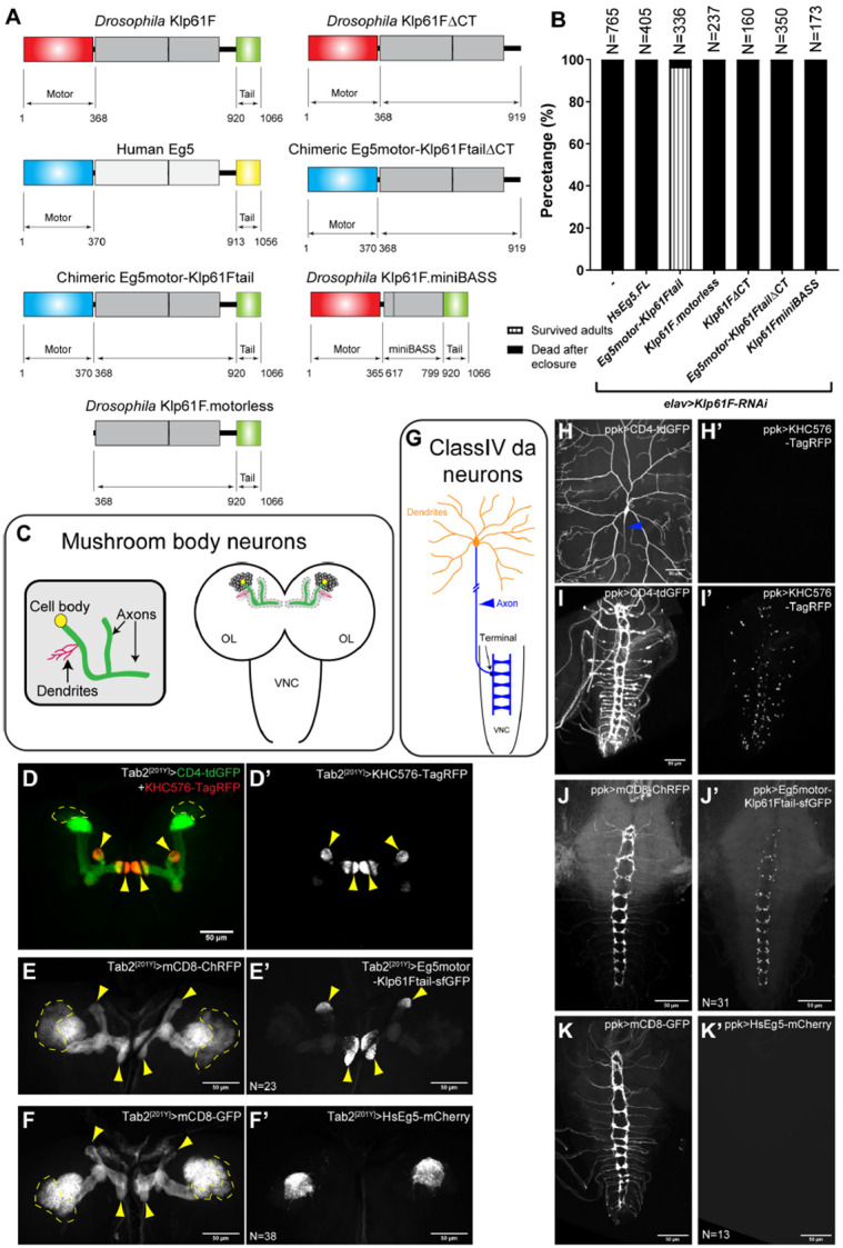
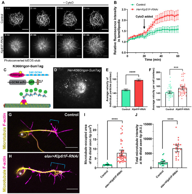
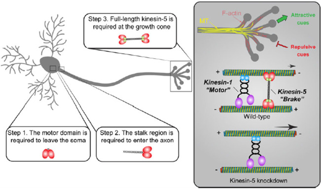
During neuronal development, neurons undergo significant microtubule reorganization to shape axons and dendrites, establishing the framework for efficient wiring of the nervous system. Previous studies from our laboratory demonstrated the key role of kinesin-1 in driving microtubule-microtubule sliding, which provides the mechanical forces necessary for early axon outgrowth and regeneration in Drosophila melanogaster. In this study, we reveal the critical role of kinesin-5, a mitotic motor, in modulating the development of postmitotic neurons. Kinesin-5, a conserved homotetrameric motor, typically functions in mitosis by sliding antiparallel microtubules apart in the spindle. Here, we demonstrate that the Drosophila kinesin-5 homolog, Klp61F, is expressed in larval brain neurons, with high levels in ventral nerve cord (VNC) neurons. Knockdown of Klp61F using a pan-neuronal driver leads to severe locomotion defects and complete lethality in adult flies, mainly due to the absence of kinesin-5 in VNC motor neurons during early larval development. Klp61F depletion results in significant axon growth defects, both in cultured and in vivo neurons. By imaging individual microtubules, we observe a significant increase in microtubule motility, and excessive penetration of microtubules into the axon growth cone in Klp61F-depleted neurons. Adult lethality and axon growth defects are fully rescued by a chimeric human-Drosophila kinesin-5 motor, which accumulates at the axon tips, suggesting a conserved role of kinesin-5 in neuronal development. Altogether, our findings show that at the growth cone, kinesin-5 acts as a brake on kinesin-1-driven microtubule sliding, preventing premature microtubule entry into the growth cone. This regulatory role of kinesin-5 is essential for precise axon pathfinding during nervous system development.


Figures

Figure 1.
Figure 1.. Kinesin-5/Klp61F is essential for neuronal function.
(A) A schematic illustration of the molecular structure of the Drosophila kinesin-5 Klp61F. (B) Survival percentage of eclosed adults in control (elavP>tdEOS-atub84B), pan-neuronal knockdown of Klp61F (elavP>Klp61F-RNAi #1, and elavP>Klp61F-RNAi #2), and pan-neuronal rescue of Klp61F depletion with RNAi-resistant full-length Klp61F (elavP>Klp61F-RNAi #1+ Klp61F (RNAi-resistant)-sfGFP). See also Video 1. (C-C’) A maximum projection of Z-stacks from the entire 3rd instar larval VNC of a Klp61F enhancer trap line, Klp61F[07012]-LacZ, stained with anti-Elav (C) and anti-βGal (C’) antibodies. Scale bar, 50 μm. See also Video 2. (D-E) A maximum projection of 2.5 μm slices of a 3rd instar larval control brain (D) and a neuronal Klp61F-depleted brain (E) stained with anti-Klp61F antibody, acquired under identical imaging conditions. Scale bars, 50 μm. (F-G’) Anti-α-tubulin (DM1α; F and G) and anti-Klp61F (F’ and G’) staining in intact (F-F’) and extracted (G-G’) Drosophila larval brain neurons. Scale bars, 5 μm. Zoom-ins of the cell body (G1) and the neurite tip (G2) are shown at the bottom.
Figure 2.
Figure 2.. Kinesin-5/Klp61F is required in early motor neurons for viability.
(A) Efficiency of the Gal4/Gal80(ts) system at permissive (18 °C) and restrictive (29 °C) temperatures. At both 18 °C and 29 °C, elavP>Klp61F-RNAi results in 100% lethality in eclosed adults. In contrast, Gal80(ts) suppresses the lethality caused by elavP>Klp61F-RNAi at 18 °C but not at 29 °C. (B) Adult survival curves of control (tubP-Gal80(ts)/+; elavP-Gal4/+) and Klp61F-RNAi (tubP-Gal80(ts)/UAS-Klp61F-RNAi #1; elavP-Gal4/+). Both samples were raised at the permissive temperature (18 °C, from embryos to adults) and then shifted to the restrictive temperature (29 °C, right after eclosure). N=84 for control and N=141 for Klp61F-RNAi. The dotted lines represent the 95% confidence intervals for each survival curve. The P value of the Gehan-Breslow-Wilcoxon test in GraphPrism is 0.8980 (not significant). (C) Temperature shifting experiments of Klp61F-RNAi flies (tubP-Gal80(ts)/UAS-Klp61F-RNAi; elavP-Gal4/+) at different development stages: 29 °C→ 18 °C, at 3rd instar larvae (L3b); 18 °C→29 °C, at 3rd instar larvae (L3b); 29 °C→ 18 °C, at 24 hours, 48 hours, 72 hours and 96 hours after egg laying (AEL). (D) Adult survival percentages with both early and late pan-neuronal expression (by elavP-Gal4), only late pan-neuronal expression (by nSyb-Gal4), and only early pan-neuronal expression (by combining elavP-Gal4 with nSyb-Gal80) of Klp61F-RNAi. (E) Adult survival percentages with both early and late motor neuron expression (by D42-Gal4), late motor neuron expression (by VGlut[OK371]-Gal4 or OK6-Gal4), or suppression of late motor neuron expression (by combining either elavP-Gal4 or D42-Gal4 with VGlut-Gal80) of Klp61F-RNAi. (F) Adult survival percentages with VNC-specific suppression (by combining elavP-Gal4 or D42-Gal4 with Tsh-Gal80) or VNC-specific expression (Tsh-Gal4) of Klp61F-RNAi. (D-F) The cartoons on the right illustrate the expression pattern of the Klp61F-RNAi (gray) at early and late stages.
Figure 3.
Figure 3.. Kinesin-5/Klp61F regulates axonal growth in culture and in vivo.
(A) Axon length measurement in cultured Drosophila larval brain neurons of control and neuronal Klp61F-RNAi. Neurons were fixed and stained with both anti-α tubulin (DM1α) and anti-Elav antibodies. The length of the longest neurite of each neuron (Elav-positive) was measured as the axon length. Scatter plots with average ± 95 % confidence intervals are shown. Unpaired t-tests with Welch's correction were performed between control and neuronal Klp61F-RNAi samples: 4 hours, p= 0.0018 (**); 1 day, p <0.0001 (****); 2 days, p= 0.0008 (***); 3 days, p=0.0080 (**). (B) A schematic illustration of the photoreceptor neurons of Drosophila 3rd instar larval eye disc and optic lobe. (C-D) Representative examples of photoreceptor neuron axonal targeting in the optic lobes of control (C) and neuronal Klp61F depletion (D). Photoreceptor neuron axons were labeled with anti-Fasciclin II antibody. Dashed lines indicate the position of the optic lobes. Scale bars, 50 μm. (E-G) Optic lobe size (E), photoreceptor neuron axon size (F), and the ratio of photoreceptor neuron axon size to the optic lobe size (G) in control and neuronal Klp61F depletion. Scatter plots with average ± 95% confidence intervals are shown. Unpaired t-tests with Welch's correction were performed between control and neuronal Klp61F-RNAi samples: optic lobe size, p <0.0001 (****); photoreceptor neuron axon size, p <0.0001 (****); the ratio of photoreceptor neuron axon size to the optic lobe size, p <0.0001 (****). (H-I) Summary of lamina (H) and medulla (I) organization phenotypes in control, neuronal Klp61F-RNAi, and rescue with RNAi-resistant full-length Drosophila Klp61F (Klp61F.FL), chimeric Eg5motor-Klp61Ftail, full-length human Eg5 (HsEg5), and Klp61F.motorless, Klp61FΔCT, chimeric Eg5motor-Klp61FtailΔCT, and Klp61F.miniBASS. Sample sizes for each genotype: control, N=85; Klp61F-RNAi, N=72; Klp61F-RNAi + Klp61F.FL, N=84; Klp61F-RNAi + Eg5motor-Klp61Ftail, N=89; Klp61F-RNAi + HsEg5, N=97; Klp61F-RNAi + Klp61F.motorless, N=86; Klp61F-RNAi + Klp61FΔCT, N=97; Klp61F-RNAi + Eg5motor-Klp61FtailΔCT, N=24; Klp61F-RNAi + Klp61F.miniBASS, N=51. All samples carried one copy of elavP-Gal4 and were stained with anti-Fasciclin II antibody to label the photoreceptor neuron axons. (J-K) Pupal motor neuron targeting in dorsal abdomen segment A5-A7 in control (J) and in elav>Klp61F-RNAi (K) 45-50 hours after pupal formation (APF). Extra branching and hyper-targeting of motor neurons in elav>Klp61F-RNAi (K) are shown with arrowheads and a bracket, respectively. Scale bars, 100 μm. (L-M) Summary of motor neuron targeting phenotypes, by the numbers of pupae (L) and the numbers of motor neuron groups (M). (N-O’) Pupal motor neuron targeting 30 hours APF in control (N) and in elav>Klp61F-RNAi (O). Extra (arrowheads) and mistargeting (bracket) neurites were seen in elav>Klp61F-RNAi compared to control. (O’) In the overlay image of the motor neuron membrane (magenta) and muscles (green, labeled with Jupiter-GFP), an axonal branch (bracket) showed aberrant targeting, deviating from its expected perpendicular orientation relative to the main axon; instead, it originated from an incorrect position and extended obliquely, eventually innervating a distant muscle (indicated by triangles). Scale bars, 20 μm. (J-K) and (N-O’) Motor neurons were labeled with membrane-targeted tdTomato driven by a motor neuron-specific driver (VGlut-LexA, LexAop-myr-tdTom); knockdown of Klp61F by RNAi was driven independently by the UAS-Gal4 system (elavP-Gal4, UASp-Klp61F-RNAi).
Figure 4.
Figure 4.. Kinesin-5/Klp61F transgene rescue and neuronal localization.
(A) Schematic illustrations of transgenes created for Klp61F-RNAi rescue experiments. All constructs, except for Human Eg5, carry silent mutations resistant to the Klp61F-RNAi (see more details in the “Molecular cloning” section of “Materials and methods”). (B) Summary of adult survival percentage in flies expressing neuronal Klp61F-RNAi and the listed transgenes. All samples carried one copy of elavP-Gal4. See also Video 1. (C) Schematic illustration of mushroom body neuron organization. Kenyon cells (the main neurons of the mushroom body) extend the dendrites at the calyx and parallel bundles of axons that are divided into either the α lobe (pointing up) or the β/γ lobes (pointing toward the center). (D-D’) Distinct compartments of axons and dendrites in the mushroom body neurons. Axon tips were labeled with a constitutively active kinesin-1 (KHC576). The mushroom body neuron membrane was labeled with CD4-tdGFP, and the dendritic-enrich calyx was labeled with the most concentrated CD4-tdGFP signal. (E-F’) Neuronal membrane labeled with mCD8-ChRFP (E-F) and localization of the transgenic chimeric motor (Eg5motor-Klp61Ftail, E’) and full-length human Eg5 (HsEg5, F’) in the mushroom body neurons. (D-F’) All transgene expressions were driven by Tab2[201Y]-Gal4. The cell bodies of Kenyon cells are indicated by dashed lines, and their axon tips are indicated by yellow arrowheads. Scale bars, 50 μm. (G) Schematic illustration of a class IV da neuron. The da neuron has elaborative dendritic arborization (orange) and extends a single unbranched axon into the VNC (blue). (H-I) Membrane labeling (with CD4-tdGFP) of the dendrites (H) and axons (I) of class IV da neurons. The proximal axon from the cell body is indicated by a blue arrowhead, and the axon goes out of focus as it extends deeper beneath the epidermis (H). (H’-I’) The constitutively active kinesin-1 (KHC576) motor is concentrated in the axon terminals at the VNC (I’) but is completely absent from the dendrites (H’). (J-K’) Neuronal membrane labeled with mCD8-ChRFP (J-K) and localization of the transgenic chimeric motor (Eg5motor-Klp61Ftail, J’) and full-length human Eg5 (HsEg5, K’) in the VNC. The dendritic localization of HsEg5 is shown in Supplemental Figure 5G-G”. (H-K’) All the transgene expressions were driven by ppk-Gal4. Scale bars, 50 μm.
Figure 5.
Figure 5.. Kinesin-5/Klp61F inhibits microtubule penetration into the periphery of hemocytes and the axonal growth cone of neurons.
(A) Microtubule penetration before and after actin fragmentation by cytochalasin D (CytoD) in control and Klp61F-depleted Drosophila primary hemocytes. CytoD was added at 20 minutes after the start of imaging. Microtubules were labeled with α-tubulin 84B tagged with a tandem dimer of EOS (tdEOS-αtub84B), and globally photoconverted from green to red by UV light. Scale bars, 5 μm. See also Videos 5 and 6. (B) Quantification of microtubule penetration into the lamellipodia in control and Klp61F-RNAi hemocytes. The microtubule intensity in the lamellipodia at 0 minutes was normalized to 1. Data points are shown as average ± SEM. Sample sizes for control and Klp61F-RNAi are N=20 and N=24, respectively. (C) Schematic illustration of the K560Rigor-SunTag system. Co-expression of (1) a human truncated kinesin-1 motor (K560) carrying a rigor mutation (E236A) with 24 copies of the GCN4 peptide, and (2) a single-chain antibody fragment (scFv) against GCN4 tagged with super-folder GFP (sfGFP) and a nuclear localization signal (NLS), result in bright dots irreversibly attached to microtubules. (D) A representative image of a K560Rigor-SunTag-expressing hemocyte. Bright dots outside the nucleus indicate the attachment points of the rigor mutant kinesin-1 motor to microtubules. The movement of these K560Rigor-SunTag dots serves as an indicator of microtubule sliding. (E-F) Quantification of K560Rigor-SunTag dot movement in control and Klp61F-RNAi hemocytes. Velocities are plotted for all particles tracked (E) or as average velocities per cell (F). The total number of particles tracked is N=8953 from 53 control cells, and N=9857 from 53 Klp61F-RNAi cells. The particles were tracked in DiaTrack 3.04. The maximum and minimum velocities were set at 500 nm/sec and 77 nm/sec, respectively. (G-H) Distribution of microtubules (by DM1α staining) and F-actin (by phalloidin staining) in control (G) and elav>Klp61F-RNAi (H) larval brain neurons 4.5 hours after plating. Asterisks indicate the cell body, and brackets indicate the axon growth cone. Scale bars, 5 μm. Both samples carried one copy of elavP-Gal4. (I-J) Microtubule distribution in the distal axon tips of control and elav>Klp61F-RNAi neurons. The distal axon tip is defined as the distal-most 5 μm of the axon containing microtubules. Microtubule distribution is shown either by the area occupied by microtubules (I) or by the total microtubule fluorescence intensity in the distal tip region (J). Sample sizes for each genotype: (I) control, N=42; elav>Klp61F-RNAi, N=47; (J) control, N=26; elav>Klp61F-RNAi, N=22. Unpaired t-tests with Welch's correction were performed between control and elav>Klp61F-RNAi: (I), p<0.0001 (****); (J), p<0.0001 (****).
Figure 6.
Figure 6.. Model of kinesin-5 function in Drosophila neurons.
Kinesin-5/Klp61F undergoes several critical steps to function properly in neurons: (1) Kinesin-5/Klp61F must leave the soma, a process that requires its motor domain, as motorless Klp61F remains exclusively localized in the neuronal cell body; (2) Kinesin-5/Klp61F must enter the axon, a process dependent on its stalk domain, since the human Eg5 motor lacking the Klp61F stalk domain is mislocalized entirely to the dendrites; (3) The full-length, active tetramer of kinesin-5/Klp61F acts as a dynamic brake on microtubule sliding driven by kinesin-1 at the axon growth cone, playing an essential role in axon pathfinding by responding to both attractive and repulsive cues.

References

    1. Baas P. W., Deitch J. S., Black M. M., Banker G. A., Polarity orientation of microtubules in hippocampal neurons: uniformity in the axon and nonuniformity in the dendrite. Proceedings of the National Academy of Sciences of the United States of America 85, 8335–8339 (1988). - PMC - PubMed
    1. Stone M.C., Roegiers F., and Rolls M.M., Microtubules Have Opposite Orientation in Axons and Dendrites of Drosophila Neurons. Molecular Biology of the Cell, 2008. 19(10): p. 4122–4129. - PMC - PubMed
    1. Yau K.W., et al., Dendrites In Vitro and In Vivo Contain Microtubules of Opposite Polarity and Axon Formation Correlates with Uniform Plus-End-Out Microtubule Orientation. J Neurosci, 2016. 36(4): p. 1071–85. - PMC - PubMed
    1. Lu W. and Gelfand V.I., Moonlighting Motors: Kinesin, Dynein, and Cell Polarity. Trends Cell Biol, 2017. - PMC - PubMed
    1. Winding M., et al., Role of kinesin-1-based microtubule sliding in Drosophila nervous system development. Proc Natl Acad Sci U S A, 2016. 113(34): p. E4985–94. - PMC - PubMed

Publication types

LinkOut - more resources