

Brain Arteriovenous Malformation Hemorrhage and Pituitary Adenoma in a COVID-19-Positive Patient
- PMID: 39314610
- PMCID: PMC11417440
- DOI: 10.7759/cureus.67644
Brain Arteriovenous Malformation Hemorrhage and Pituitary Adenoma in a COVID-19-Positive Patient
Abstract
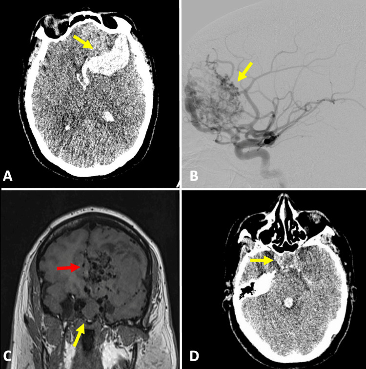
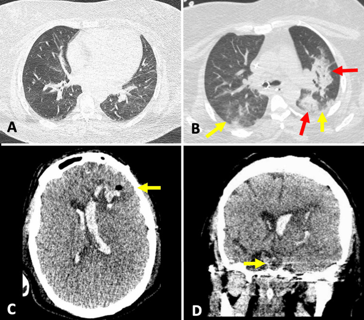
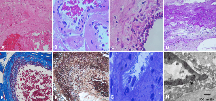
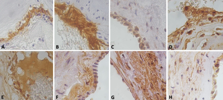
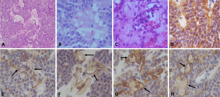
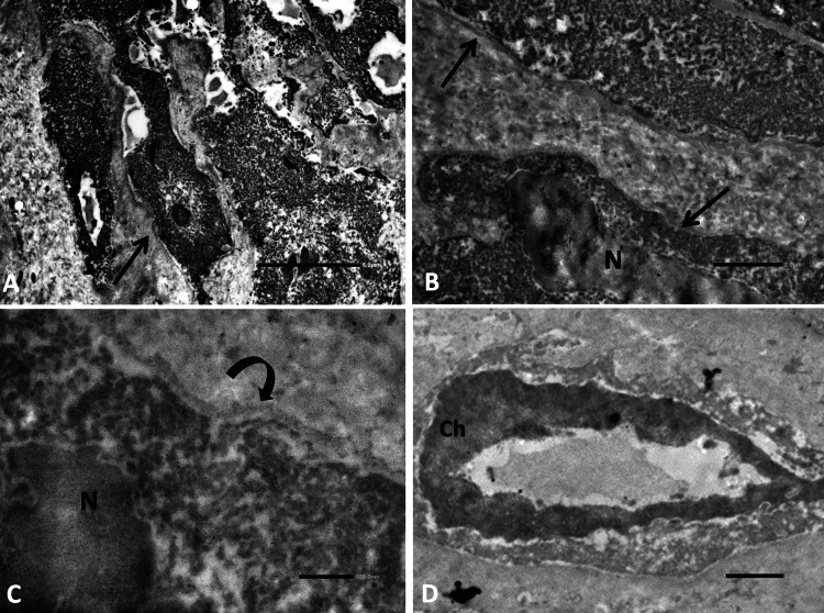
Brain arteriovenous malformations (AVMs) are usually asymptomatic. They can cause intense pain or bleeding or lead to other serious medical problems. We present a rare case of a woman who presented with a severe headache and was brought to the emergency service for an intracerebral hemorrhage due to a ruptured AVM. During the surgery, a sellar mass was identified that was also resected. AVM showed vasculitis, endarteritis, endothelial damage, leukocyte plug, and damage to the vessel wall with fragmentation of the collagen and actin filaments. The sellar mass showed a non-functioning pituitary adenoma with hemorrhagic foci and necrosis as well as a proteinaceous vs. lipid material deposition with minimal vascular changes such as endothelial hyperplasia with minimal vasculitis and hyperplasia of reticular stellate cells, with positive glial fibrillary acidic protein (GFAP), which expressed low expression of luteinizing hormone (LH) and follicle-stimulating hormone (FSH), IL6, IL10, IL17, tumor necrosis factor-alpha (TNFa), HIF1a, factor VIII (FVIII), platelet-derived growth factor (PDGF), vascular endothelial growth factor (VEGF), and VEGF receptor 2 (VEGFR2). The patient's polymerase chain reaction COVID-19 test was positive, and she died three days after the surgery procedure. In our knowledge of COVID-19 brain lesions and in the literature review, this was a rare case of a double pathology associated with COVID-19 infection characterized by rupture of the AVM with hemorrhages and brain infarcts associated with endarteritis, vessel wall injuries, and pituitary apoplexy.
Keywords: brain arteriovenous malformation; cerebrovascular disease; covid-19; pituitary adenoma; pituitary apoplexy.
Copyright © 2024, Nathal et al.
Conflict of interest statement
Human subjects: Consent was obtained or waived by all participants in this study. Conflicts of interest: In compliance with the ICMJE uniform disclosure form, all authors declare the following: Payment/services info: All authors have declared that no financial support was received from any organization for the submitted work. Financial relationships: All authors have declared that they have no financial relationships at present or within the previous three years with any organizations that might have an interest in the submitted work. Other relationships: All authors have declared that there are no other relationships or activities that could appear to have influenced the submitted work.
Figures
Similar articles
-
Deciphering the vascular labyrinth: role of microRNAs and candidate gene SNPs in brain AVM development - literature review.Neurol Res. 2020 Dec;42(12):1043-1054. doi: 10.1080/01616412.2020.1796380. Epub 2020 Jul 28. Neurol Res. 2020. PMID: 32723034 Review.
-
Reductions in brain pericytes are associated with arteriovenous malformation vascular instability.J Neurosurg. 2018 Dec 1;129(6):1464-1474. doi: 10.3171/2017.6.JNS17860. Epub 2018 Jan 5. J Neurosurg. 2018. PMID: 29303444 Free PMC article.
-
Fatal ruptured occult arteriovenous malformation in a young adult: An autopsy case report.Surg Neurol Int. 2022 Jul 1;13:284. doi: 10.25259/SNI_427_2022. eCollection 2022. Surg Neurol Int. 2022. PMID: 35855123 Free PMC article.
-
Delayed treatment of ruptured brain AVMs: is it ok to wait?J Neurosurg. 2018 Apr;128(4):999-1005. doi: 10.3171/2017.1.JNS16745. Epub 2017 Jul 7. J Neurosurg. 2018. PMID: 28686111
-
Sudden death in custody due to pituitary apoplexy during long restriction in a sitting position: a case report and review of the literature.J Forensic Leg Med. 2013 Oct;20(7):812-5. doi: 10.1016/j.jflm.2013.06.012. Epub 2013 Aug 6. J Forensic Leg Med. 2013. PMID: 24112326 Review.
References
-
- Intracerebral hemorrhage in young people: analysis of risk factors, location, causes, and prognosis. Ruíz-Sandoval JL, Cantú C, Barinagarrementeria F. Stroke. 1999;30:537–541. - PubMed
-
- A systematic review of the frequency and prognosis of arteriovenous malformations of the brain in adults. Al-Shahi R, Warlow C. Brain. 2001;124:1900–1926. - PubMed
-
- The natural history and predictive features of hemorrhage from brain arteriovenous malformations. da Costa L, Wallace MC, Ter Brugge KG, O'Kelly C, Willinsky RA, Tymianski M. Stroke. 2009;40:100–105. - PubMed
Publication types
LinkOut - more resources
Full Text Sources
Miscellaneous