Case Reports
. 2024 Sep 13;19(12):5844-5848.
doi: 10.1016/j.radcr.2024.08.124. eCollection 2024 Dec.

Unusual presentation of a splenic infarction

Affiliations
Case Reports

Unusual presentation of a splenic infarction

Kolos K Nagy et al. Radiol Case Rep. .

Abstract

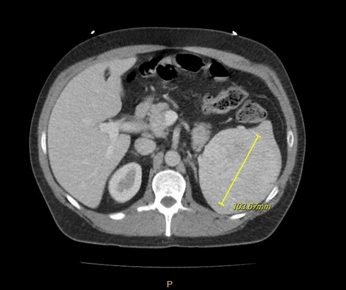
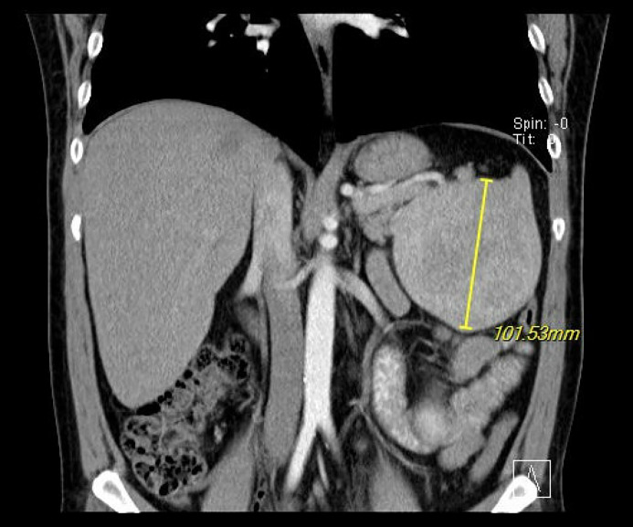
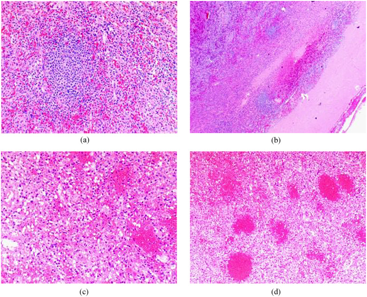
A 41-year-old male with a history of tobacco and alcohol use presented to our clinic for a follow up of an incidentally diagnosed splenic mass. The patient was sent for further diagnostic evaluation, and computed tomography showed a large splenic mass with heterogenous enhancement raising concern for neoplasm. Due to the uncertain nature of the splenic lesion and high complication rate of percutaneous splenic biopsy, a splenectomy was performed. The specimen was sent to pathology, and the report favored neoplasm but was inconclusive. The samples were sent to another institution for a consult, where the patient's spleen was determined to be the result of a previously suffered hemorrhagic infarct. This case demonstrates the difficulty of diagnosing splenic lesions using diagnostic imaging and the discrepancy that may occur between radiology and pathology assessments. In the setting of uncertain pathology, the removal of what might be a functional spleen is often preferred over a percutaneous biopsy due to concerns of intraabdominal bleeding and tumor dissemination.

Keywords: Case report; Splenectomy; Splenic infarction; Splenic lesion; Splenic neoplasm.


Figures

Fig 1:
Fig. 1
Axial view CT w/ contrast of splenic mass.
Fig 2:
Fig. 2
Coronal view CT w/ contrast of splenic mass.
Fig 3
Fig. 3
(A) Low power (100x) view of normal spleen parenchyma. (B) Low power (200x) view of subcapsular area showing infarction. (C) Intermediate (200x) view of infarction. (D) High power (400x) view showing infarction.
Fig 4:
Fig. 4
Axial view CT w/ contrast demonstrating enlarged splenic vein.

References

    1. Saboo SS, Krajewski KM, O’Regan KN, Giardino A, Brown JR, Ramaiya N, et al. Spleen in haematological malignancies: spectrum of imaging findings. Br J Radiol. 2012;85(1009):81–92. doi: 10.1259/bjr/31542964. - DOI - PMC - PubMed
    1. Kim N, Auerbach A, Manning MA. Algorithmic approach to the splenic lesion based on radiologic-pathologic correlation. Radiographics. 2022;42(3):683–701. doi: 10.1148/rg.210071. - DOI - PubMed
    1. Ozakin E, Cetinkaya O, Baloglu Kaya F, Acar N, Cevik AA. A rare cause of acute abdominal pain: splenic infarct (case series) Turk J Emerg Med. 2016;15(2):96–99. doi: 10.5505/1304.7361.2015.16769. - DOI - PMC - PubMed
    1. Barrak D, Ramly EP, Chouillard E, Khoury M. Chronic spontaneous idiopathic spleen hematoma presenting as a large cystic tumor: a case report with review of the literature. J Surg Case Rep. 2014;2014(6):6–9. doi: 10.1093/jscr/rju060. - DOI - PMC - PubMed
    1. Hiyama K, Kirino I, Fukui Y, Terashima H. Two cases of splenic neoplasms with differing imaging findings that required laparoscopic resection for a definitive diagnosis. Int J Surg Case Rep. 2022;93 doi: 10.1016/j.ijscr.2022.107023. - DOI - PMC - PubMed

Publication types

LinkOut - more resources