Functional Neural Networks in Human Brain Organoids
- PMID: 39314749
- PMCID: PMC11418062
- DOI: 10.34133/bmef.0065
Functional Neural Networks in Human Brain Organoids
Abstract
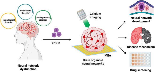
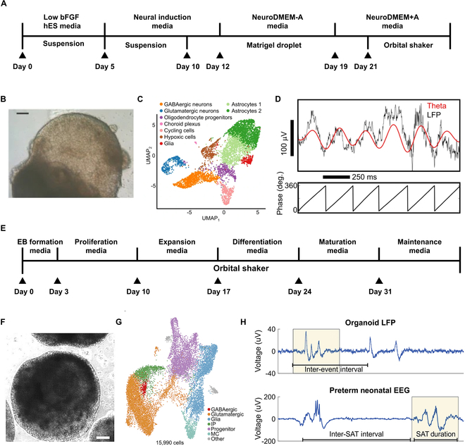
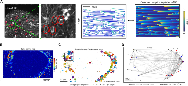
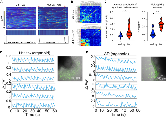
Human brain organoids are 3-dimensional brain-like tissues derived from human pluripotent stem cells and hold promising potential for modeling neurological, psychiatric, and developmental disorders. While the molecular and cellular aspects of human brain organoids have been intensively studied, their functional properties such as organoid neural networks (ONNs) are largely understudied. Here, we summarize recent research advances in understanding, characterization, and application of functional ONNs in human brain organoids. We first discuss the formation of ONNs and follow up with characterization strategies including microelectrode array (MEA) technology and calcium imaging. Moreover, we highlight recent studies utilizing ONNs to investigate neurological diseases such as Rett syndrome and Alzheimer's disease. Finally, we provide our perspectives on the future challenges and opportunities for using ONNs in basic research and translational applications.
Copyright © 2024 Longjun Gu et al.
Conflict of interest statement
Competing interests: The authors declare that they have no competing interests.
Figures
Similar articles
-
Challenges in Modeling Human Neural Circuit Formation via Brain Organoid Technology.Front Cell Neurosci. 2020 Dec 3;14:607399. doi: 10.3389/fncel.2020.607399. eCollection 2020. Front Cell Neurosci. 2020. PMID: 33362473 Free PMC article. Review.
-
Advances in Central Nervous System Organoids: A Focus on Organoid-Based Models for Motor Neuron Disease.Tissue Eng Part C Methods. 2021 Mar;27(3):213-224. doi: 10.1089/ten.TEC.2020.0337. Epub 2021 Mar 3. Tissue Eng Part C Methods. 2021. PMID: 33446055 Review.
-
3D brain Organoids derived from pluripotent stem cells: promising experimental models for brain development and neurodegenerative disorders.J Biomed Sci. 2017 Aug 20;24(1):59. doi: 10.1186/s12929-017-0362-8. J Biomed Sci. 2017. PMID: 28822354 Free PMC article. Review.
-
Application of Human Brain Organoids-Opportunities and Challenges in Modeling Human Brain Development and Neurodevelopmental Diseases.Int J Mol Sci. 2023 Aug 7;24(15):12528. doi: 10.3390/ijms241512528. Int J Mol Sci. 2023. PMID: 37569905 Free PMC article. Review.
-
Studying Human Neurodevelopment and Diseases Using 3D Brain Organoids.J Neurosci. 2020 Feb 5;40(6):1186-1193. doi: 10.1523/JNEUROSCI.0519-19.2019. J Neurosci. 2020. PMID: 32024767 Free PMC article. Review.
Cited by
-
Engineering blood-brain barrier microphysiological systems to model Alzheimer's disease monocyte penetration and infiltration.Biomater Sci. 2025 Jun 25;13(13):3650-3661. doi: 10.1039/d5bm00204d. Biomater Sci. 2025. PMID: 40391576
-
Self-Organizing Neural Networks in Organoids Reveal Principles of Forebrain Circuit Assembly.bioRxiv [Preprint]. 2025 May 2:2025.05.01.651773. doi: 10.1101/2025.05.01.651773. bioRxiv. 2025. PMID: 40654898 Free PMC article. Preprint.
-
Beyond Structure: Next-Generation Electrophysiological Platforms for Functional Brain Organoids.Int J Stem Cells. 2025 Aug 30;18(3):215-236. doi: 10.15283/ijsc25056. Epub 2025 Jul 31. Int J Stem Cells. 2025. PMID: 40739714 Free PMC article. Review.
-
Navigating Brain Organoid Maturation: From Benchmarking Frameworks to Multimodal Bioengineering Strategies.Biomolecules. 2025 Aug 4;15(8):1118. doi: 10.3390/biom15081118. Biomolecules. 2025. PMID: 40867563 Free PMC article. Review.
-
Vascular network-inspired diffusible scaffolds for engineering functional midbrain organoids.Cell Stem Cell. 2025 May 1;32(5):824-837.e5. doi: 10.1016/j.stem.2025.02.010. Epub 2025 Mar 17. Cell Stem Cell. 2025. PMID: 40101722 Free PMC article.
References
-
- Bressler SL, Menon V. Large-scale brain networks in cognition: Emerging methods and principles. Trends Cogn Sci. 2010;14(6):277–290. - PubMed
-
- Neves G, Cooke SF, TVP Bliss. Synaptic plasticity, memory, and the hippocampus: A neural network approach to causality. Nat Rev Neurosci. 2008;9(1):65–75. - PubMed
Publication types
LinkOut - more resources
Full Text Sources