

This is a preprint.
Computed tomography radiomics-based cross-sectional detection of mandibular osteoradionecrosis in head and neck cancer survivors
- PMID: 39314948
- PMCID: PMC11419222
- DOI: 10.1101/2024.09.11.24313485
Computed tomography radiomics-based cross-sectional detection of mandibular osteoradionecrosis in head and neck cancer survivors
Update in
-
Computed tomography radiomics-based cross-sectional detection of mandibular osteoradionecrosis in head and neck cancer survivors.Oral Oncol. 2025 Aug;167:107337. doi: 10.1016/j.oraloncology.2025.107337. Epub 2025 Jun 13. Oral Oncol. 2025. PMID: 40516152 Free PMC article.
Abstract
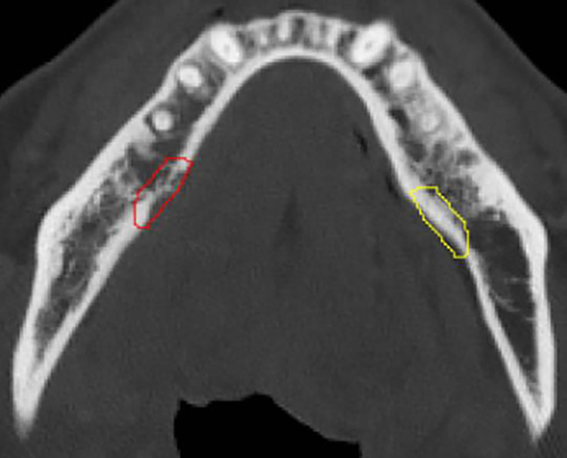
Purpose: This study aims to identify radiomic features extracted from contrast-enhanced CT scans that differentiate osteoradionecrosis (ORN) from normal mandibular bone in patients with head and neck cancer (HNC) treated with radiotherapy (RT).
Materials and methods: Contrast-enhanced CT (CECT) images were collected for 150 patients (80% train, 20% test) with confirmed ORN diagnosis at The University of Texas MD Anderson Cancer Center between 2008 and 2018. Using PyRadiomics, radiomic features were extracted from manually segmented ORN regions and the corresponding automated control regions, the later defined as the contralateral healthy mandible region. A subset of pre-selected features was obtained based on correlation analysis (r > 0.95) and used to train a Random Forest (RF) classifier with Recursive Feature Elimination. Model explainability SHapley Additive exPlanations (SHAP) analysis was performed on the 20 most important features identified by the trained RF classifier.
Results: From a total of 1316 radiomic features extracted, 810 features were excluded due to high collinearity. From a set of 506 pre-selected radiomic features, the optimal subset resulting on the best discriminative accuracy of the RF classifier consisted of 67 features. The RF classifier was well calibrated (Log Loss 0.296, ECE 0.125) and achieved an accuracy of 88% and a ROC AUC of 0.96. The SHAP analysis revealed that higher values of Wavelet-LLH First-order Mean and Median were associated with ORN of the jaw (ORNJ). Conversely, higher Exponential GLDM Dependence Entropy and lower Square First-order Kurtosis were more characteristic of normal mandibular tissue.
Conclusion: This study successfully developed a CECT-based radiomics model for differentiating ORNJ from healthy mandibular tissue in HNC patients after RT. Future work will focus on the detection of subclinical ORNJ regions to guide earlier interventions.
Conflict of interest statement
Conflict of Interest Statement: Dr. Fuller has received unrelated direct industry grant/in-kind support, honoraria, and travel funding from Elekta AB; honoraria, and travel funding from Philips Medical Systems; and honoraria, and travel funding from Varian/Siemens Healthineers. Dr. Fuller has unrelated licensing/royalties from Kallisio, Inc. Dr. Sandulache is a consultant for, and equity holder in, Femtovox Inc (unrelated to current work).
Figures


References
Publication types
Grants and funding
LinkOut - more resources
Full Text Sources
Miscellaneous