

Characterization of cortical volume and whole-brain functional connectivity in Parkinson's disease patients: a MRI study combined with physiological aging brain changes
- PMID: 39315074
- PMCID: PMC11418396
- DOI: 10.3389/fnins.2024.1451948
Characterization of cortical volume and whole-brain functional connectivity in Parkinson's disease patients: a MRI study combined with physiological aging brain changes
Abstract
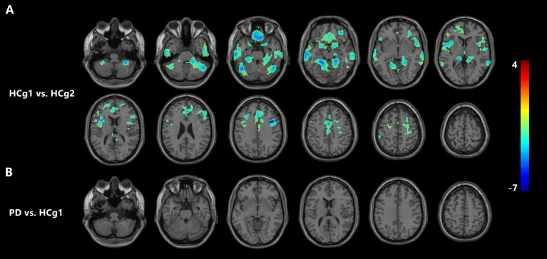
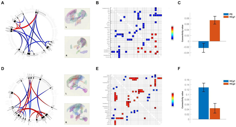
This study employed multiple MRI features to comprehensively evaluate the abnormalities in morphology, and functionality associated with Parkinson's disease (PD) and distinguish them from normal physiological changes. For investigation purposes, three groups: 32 patients with PD, 42 age-matched healthy controls (HCg1), and 33 young and middle-aged controls (HCg2) were designed. The aim of the current study was to differentiate pathological cortical changes in PD from age-related physiological cortical volume changes. Integrating these findings with functional MRI changes to characterize the effects of PD on whole-brain networks. Cortical volumes in the bilateral temporal lobe, frontal lobe, and cerebellum were significantly reduced in HCg1 compared to HCg2. Although no significant differences in cortical volume were observed between PD patients and HCg1, the PD group exhibited pronounced abnormalities with significantly lower mean connectivity values compared to HCg1. Conversely, physiological functional changes in HCg1 showed markedly higher mean connectivity values than in HCg2. By integrating morphological and functional assessments, as well as network characterization of physiological aging, this study further delineates the distinct characteristics of pathological changes in PD.
Keywords: Parkinson’s disease; cortex; fMRI; functional connectivity; morphology.
Copyright © 2024 Wang, Chen, Zhang, Gao, Gou and Lei.
Conflict of interest statement
The authors declare that the research was conducted in the absence of any commercial or financial relationships that could be construed as a potential conflict of interest.
Figures
References
-
- Alloza C., Cox S. R., Blesa Cábez M., Redmond P., Whalley H. C., Ritchie S. J., et al. (2018). Polygenic risk score for schizophrenia and structural brain connectivity in older age: a longitudinal connectome and tractography study. NeuroImage 183, 884–896. doi: 10.1016/j.neuroimage.2018.08.075 - DOI - PMC - PubMed
LinkOut - more resources
Full Text Sources