. 2024 Sep 9:11:1440769.
doi: 10.3389/fvets.2024.1440769. eCollection 2024.

Development of a RPA-CRISPR/Cas12a based rapid visual detection assay for Porcine Parvovirus 7

Affiliations

Development of a RPA-CRISPR/Cas12a based rapid visual detection assay for Porcine Parvovirus 7

Shubo Wen et al. Front Vet Sci. .

Abstract

Introduction: Porcine Parvovirus (PPV) is a significant pathogen in the pig industry, with eight genotypes, including PPV7, identified since its emergence in 2016. Co-infections with viruses such as Porcine Circovirus 2 (PCV2) and Porcine Reproductive and Respiratory Syndrome Virus (PRRSV) pose serious risks to swine health. Thus, there is an urgent need for rapid, sensitive, and specific detection methods suitable for use in field settings or laboratories with limited resources.

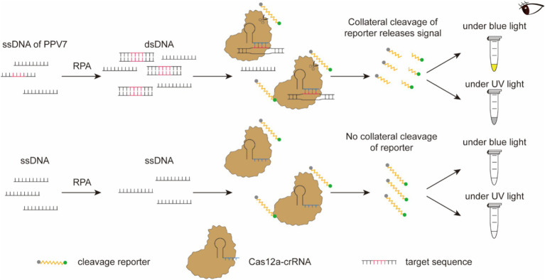
Methods: We developed a CRISPR/Cas12a-based assay combined with recombinase polymerase amplification (RPA) for the rapid detection of PPV7. Specific RPA primers and five CRISPR RNAs (crRNAs) were designed to target a highly conserved region within the NS1 gene of PPV7. Optimization of crRNA and single-stranded DNA (ssDNA) concentrations was performed to enhance the assay's performance.

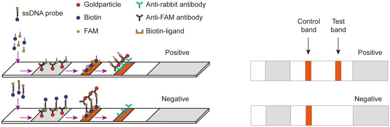
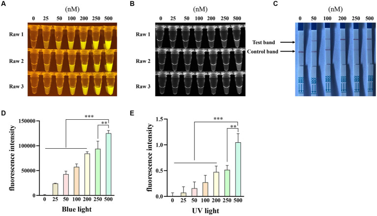
Results: CrRNA optimization identified crRNA-05 as the optimal candidate for Cas12a-based detection of PPV7, as all synthesized crRNAs demonstrated similar performance. The optimal crRNA concentration was determined to be 200 nM, yielding consistent results across tested concentrations. For ssDNA optimization, the strongest fluorescence signal was achieved with 500 nM of the FAM-BHQ ssDNA receptor. The assay showed a minimal detection limit of 100copies/μl for PPV7, confirmed through fluorescence and lateral flow detection methods. Specificity testing indicated that only PPV7 DNA samples returned positive results, confirming the assay's accuracy. In tests of 50 lung tissue samples from diseased pigs, the RPA-Cas12a assay identified 29 positive samples (58%), surpassing the 22 positive samples (44%) detected by conventional PCR. This highlights the RPA-Cas12a method's enhanced detection capability and its potential utility in clinical surveillance and management of PPV7 in swine populations.

Discussion: The RPA-Cas12a assay effectively detects PPV7 in clinical samples, enhancing disease surveillance and control in pigs. Its adaptability to resource-limited settings significantly improves PPV7 management and prevention strategies, thereby supporting the overall health and development of the pig industry.

Keywords: CRISPR/Cas12a; PCR; Porcine Parvovirus 7; detection; visual.


Conflict of interest statement

AL was employed by Guangzhou Yitun Pig Industry Co. Ltd. The remaining authors declare that the research was conducted in the absence of any commercial or financial relationships that could be construed as a potential conflict of interest.

Figures

Figure 1
Figure 1
The primer pair designed for RPA amplification was algined with the NS1 gene of ten randomly selected PPV7 strains from GenBank using MEGA 7.0 software. The primer sequences are indicated with solid black boxes.
Figure 2
Figure 2
Schematic diagram of the RPA-CRISPR/Cas12a-based nucleic acid detection platform. Following amplification, the RPA product is employed for the detection assay using CRISPR/Cas12a. An ssDNA tagged with a quenched green fluorescent molecule is introduced for visual detection purposes. When the target molecule is present in the system, Cas12a cleaves the ssDNA, triggering a visible green fluorescence signal. In the absence of virus-derived dsDNA, the ssDNA remains uncleaved, resulting in no fluorescence signal generation. The detection outcomes can be directly observed by the naked eyes under blue light or UV light after an incubation period of approximately 15 min.
Figure 3
Figure 3
Schematic diagram of Lateral flow dipstick detection. Gold nanoparticles, conjugated with an anti-FAM antibody, were positioned on the binding pad. Streptavidin and IgG were fixed onto the NC membrane, serving as a control line for specific biotin binding and a test line for specifically binding to an anti-FAM antibody, respectively. The FAM-Biotin ssDNA reporter selectively adhered to the gold nanoparticles to create a complex due to the recognition of FAM by the anti-FAM antibody on the gold nanoparticles. When the Cas12a did not degrade the ssDNA, this complex bound to streptavidin at the control line. Conversely, when the ssDNA was degraded, leading to the release of Biotin from the complex, the complex was able to transit past the control line and bind to the IgG antibody at the test line.
Figure 4
Figure 4
Selection of the optimal crRNA for PPV7 detection by CRISPR/Cas12a-based assay. The CRISPR/Cas12a based fluorescence detection assay using 5 different crRNAs under blue light (A) and UV light (B). Lateral flow strip assay using 5 crRNAs (C). Fluorescent intensity was calculated by Image J software (D,E). DNA template represents conserved sequence of NS1 in pMD18T-NS1 plasmid; no. 1–5 represent crRNA-01-crRNA-05, respectively. NC: negative control (ddH2O). All the experiments were performed three time. Values are presented as means ± s.d. (error bars) (n = 3 replicates; * p < 0.05, ** p < 0.01, *** p < 0.001 between samples, two-sample t-test).
Figure 5
Figure 5
Optimization of the crRNA concentration. The CRISPR/Cas12a based fluorescence detection assay with different concentrations of crRNA under blue light (A) and UV light (B). Lateral flow strip assay with different concentrations of crRNA (C). Fluorescent intensity was calculated by Image J software (D,E). NC: negative control (ddH2O). All the experiments were performed three time. Values are presented as means ± s.d. (error bars) (n = 3 replicates; * p < 0.05, ** p < 0.01, *** p < 0.001 between samples, two-sample t-test).
Figure 6
Figure 6
Optimization of the concentration of single-stranded DNA-fluorescent quencher (ssDNA-FQ) reporters and biotin for Porcine Parvovirus 7 (PPV7) detection by a CRISPR/Cas12a-based assay. The CRISPR/Cas12a based fluorescence detection assay with varying concentrations of ssDNA-FQ reporters under blue light (A) and UV light (B). Lateral flow strip assay conducted with different concentrations of ssDNA-biotin (C). NC: negative control (ddH2O). All the experiments were performed three time. Values are presented as means ± s.d. (error bars) (n = 3 replicates; * p < 0.05, ** p < 0.01, *** p < 0.001 between samples, two-sample t-test). Fluorescent intensity was calculated by Image J software (D and E).
Figure 7
Figure 7
Sensitivity of RPA-CRISPR/Cas12a-based detection for PPV7. Serial 10-fold dilutions of PPV7 pUC57-NS1 plasmids (108–100 copies) were detected using the RPA-CRISPR/Cas12a-based fluorescence detection assay (A), or (106–100 copies) were detected using the lateral-flow strip assay (B). Additionally, the sensitivity of the traditional PCR detection method used in this study was evaluated using the same concentration gradient of pUC57-NS1 (C). No. 1–9 represents plasmids with concentrations of 108–100 copies/μl in (A,C), and no. 1–7 represents plasmids with concentrations of 106–100 copies/μl in (B). NC: negative control (ddH2O).
Figure 8
Figure 8
Specificity of the RPA-CRISPR/Cas12a-based detection for PPV7. The specificity analysis of the RPA-CRISPR/Cas12a-based fluorescence detection assay was carried out using the viral genomic DNA of a PPV2-PPV6 mixture, PRV, PCV2, PCV3, and complementary DNA (cDNA) of PRRSV in the fluorescence detection assay (A), in the lateral-flow strip assay (B) and in the traditional PCR detection method (C). No. 1–2 represents PPV7 pUC57-NS1 plasmids, while 3–7 represent the viral genomic DNA of the PPV2-PPV6 mixture, PRV, PCV2, PCV3, and cDNA of PRRSV. NC: negative control (ddH2O).

Similar articles

References

    1. Streck AF, Canal CW, Truyen U. Molecular epidemiology and evolution of porcine parvoviruses. Infect Genet Evol. (2015) 36:300–6. doi: 10.1016/j.meegid.2015.10.007, PMID: - DOI - PubMed
    1. Palinski RM, Mitra N, Hause BM. Discovery of a novel Parvovirinae virus, porcine parvovirus 7, by metagenomic sequencing of porcine rectal swabs. Virus Genes. (2016) 52:564–7. doi: 10.1007/s11262-016-1322-1, PMID: - DOI - PubMed
    1. Guo Y, Yan G, Chen S, Han H, Li J, Zhang H, et al. . Identification and genomic characterization of a novel porcine parvovirus in China. Front Vet Sci. (2022) 9:1009103. doi: 10.3389/fvets.2022.1009103, PMID: - DOI - PMC - PubMed
    1. Opriessnig T, Halbur PG. Concurrent infections are important for expression of porcine circovirus associated disease. Virus Res. (2012) 164:20–32. doi: 10.1016/j.virusres.2011.09.014, PMID: - DOI - PMC - PubMed
    1. Allan GM, Kennedy S, McNeilly F, Foster JC, Ellis JA, Krakowka SJ, et al. . Experimental reproduction of severe wasting disease by co-infection of pigs with porcine circovirus and porcine parvovirus. J Comp Pathol. (1999) 121:1–11. doi: 10.1053/jcpa.1998.0295, PMID: - DOI - PubMed

LinkOut - more resources