

The markers of the predictive DNA test for canine hip dysplasia may have a stronger relationship with elbow dysplasia
- PMID: 39315210
- PMCID: PMC11417230
- DOI: 10.1016/j.heliyon.2024.e37716
The markers of the predictive DNA test for canine hip dysplasia may have a stronger relationship with elbow dysplasia
Abstract
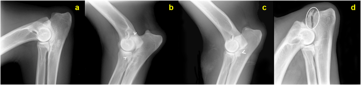
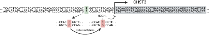
Canine hip and elbow dysplasias, which are prevalent orthopedic conditions rooted in developmental and hereditary factors are yet to be comprehensively assessed. This study aimed to address this gap by exploring the prognostic significance of five markers linked to canine hip dysplasia using available genome-wide association studies (GWAS) data. The influence of these markers on both hip and elbow dysplasia was examined in dogs exposed to standardized environmental conditions. We made a groundbreaking discovery using custom primers, qPCR assays, and evaluation of fluorescent resonance energy transfer (FRET) probes. Three specific SNPs previously associated with the risk of canine hip dysplasia demonstrated a potentially stronger correlation with elbow dysplasia. Notably, the SNP at nucleotide position 22691322, located near the canine CHST3 gene, displayed significance as a marker in multivariable logistic regression analysis. Surprisingly, none of the initially targeted SNPs showed a direct association with hip dysplasia. The genomic positions of these SNPs reside within a region conserved across mammals. In silico analyses suggested that the relevant variant might be positioned in a region linked to bone and muscle structures. Our findings revealed a remarkable relationship between SNP2 genotypes and methylation patterns, shedding light on the underlying mechanism that partially explains the genotype-phenotype correlation in canine CHST3. These groundbreaking findings offer essential insights for future, more extensive investigations into canine orthopedic health. This research significantly contributes to our understanding of the molecular foundations of hip and elbow dysplasia in dogs by charting a course for advancements in veterinary medicine and the overall well-being of canine companions.
Keywords: Dog; Elbow dysplasia; FRET probes; Hip dysplasia; In silico; SNP.
© 2024 The Authors.
Conflict of interest statement
The authors declare that they have no known competing financial interests or personal relationships that could have appeared to influence the work reported in this paper.
Figures


Similar articles
-
A genetic predictive model for canine hip dysplasia: integration of Genome Wide Association Study (GWAS) and candidate gene approaches.PLoS One. 2015 Apr 13;10(4):e0122558. doi: 10.1371/journal.pone.0122558. eCollection 2015. PLoS One. 2015. PMID: 25874693 Free PMC article.
-
Identification of quantitative trait loci (QTL) for canine hip dysplasia and canine elbow dysplasia in Bernese mountain dogs.PLoS One. 2012;7(11):e49782. doi: 10.1371/journal.pone.0049782. Epub 2012 Nov 26. PLoS One. 2012. PMID: 23189162 Free PMC article.
-
Across-breed genetic investigation of canine hip dysplasia, elbow dysplasia, and anterior cruciate ligament rupture using whole-genome sequencing.Front Genet. 2022 Dec 2;13:913354. doi: 10.3389/fgene.2022.913354. eCollection 2022. Front Genet. 2022. PMID: 36531249 Free PMC article.
-
Selection against canine hip dysplasia: success or failure?Vet J. 2011 Aug;189(2):160-8. doi: 10.1016/j.tvjl.2011.06.014. Epub 2011 Jul 2. Vet J. 2011. PMID: 21727013 Review.
-
Canine elbow dysplasia: aetiopathogenesis and current treatment recommendations.Vet J. 2013 Apr;196(1):12-9. doi: 10.1016/j.tvjl.2012.11.009. Epub 2012 Dec 23. Vet J. 2013. PMID: 23266351 Review.
Cited by
-
AI-driven techniques for detection and mitigation of SARS-CoV-2 spread: a review, taxonomy, and trends.Clin Exp Med. 2025 Jun 14;25(1):204. doi: 10.1007/s10238-025-01753-5. Clin Exp Med. 2025. PMID: 40515956 Free PMC article. Review.
-
The interaction between a leflunomide-response methylation site (cg17330251) and variant (rs705379) on response to leflunomide in patients with rheumatoid arthritis.Front Pharmacol. 2025 Mar 20;16:1499723. doi: 10.3389/fphar.2025.1499723. eCollection 2025. Front Pharmacol. 2025. PMID: 40183079 Free PMC article.
References
-
- Mäki K., Janss L., Groen A., Liinamo A., Ojala M. An indication of major genes affecting hip and elbow dysplasia in four Finnish dog populations. Heredity. 2004;92(5):402–408. - PubMed
-
- Kirberger R., Fourie S. Elbow dysplasia in the dog: pathophysiology, diagnosis and control. J. S. Afr. Vet. Assoc. 1998;69(2):43–54. - PubMed
-
- Michelsen J. Canine elbow dysplasia: aetiopathogenesis and current treatment recommendations. Vet. J. 2013;196(1):12–19. - PubMed
-
- Todhunter R.J., Bliss S., Casella G., et al. Genetic structure of susceptibility traits for hip dysplasia and microsatellite informativeness of an outcrossed canine pedigree. J. Hered. 2003;94(1):39–48. - PubMed
-
- Atalay S., Kök S. The effects of polymorphisms in the CX3CR1 gene on the development of canine hipdysplasia. Turk. J. Vet. Anim. Sci. 2022;46(1):74–87.
LinkOut - more resources
Full Text Sources
Miscellaneous
