. 2024 Sep 5;10(18):e37512.
doi: 10.1016/j.heliyon.2024.e37512. eCollection 2024 Sep 30.

Pseudomonas aeruginosa exotoxin A as a novel allergen induced Non-TH2 inflammation in a murine model of steroid-insensitive asthma

Affiliations

Pseudomonas aeruginosa exotoxin A as a novel allergen induced Non-TH2 inflammation in a murine model of steroid-insensitive asthma

Huancheng Xie et al. Heliyon. .

Abstract

Background: Despite the immediate in vivo occurrence of anaphylactic and allergic reactions following treatment with Pseudomonas aeruginosa exotoxin A (PEA)-based immunotoxins, the immunological role of PEA in asthma pathogenesis remains unclear.

Objective: This study investigated the allergenic potential of PEA and the specific type of asthma induced.

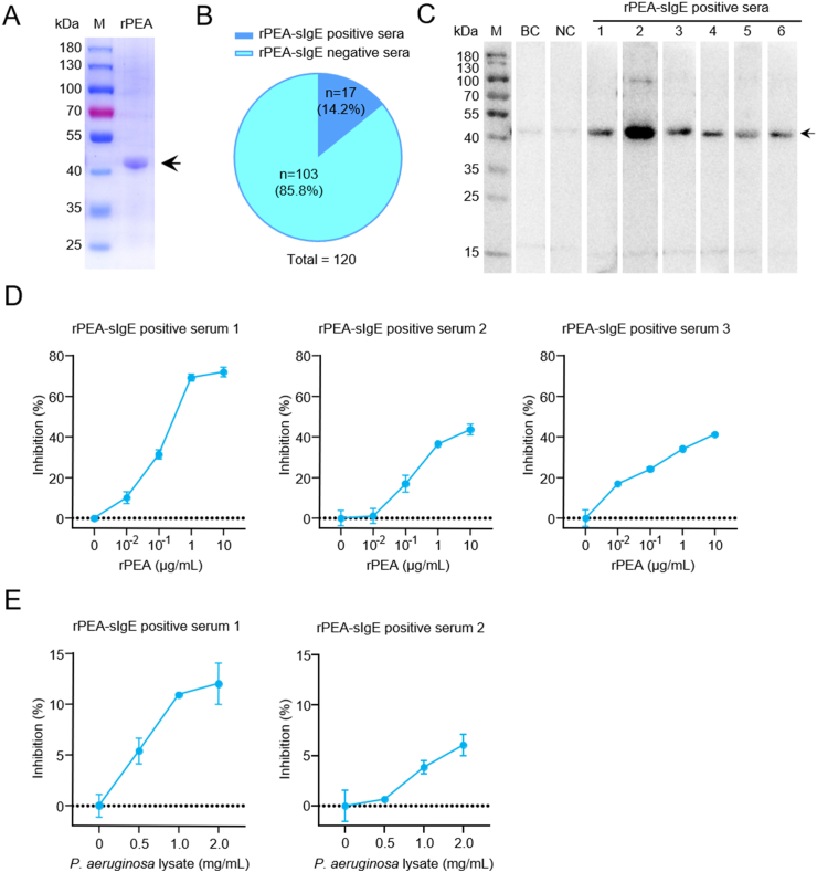
Methods: Recombinant PEA (rPEA) lacking domain Ia (to eliminate non-specific cytotoxicity) was expressed, purified, and employed to detect serum PEA-specific IgE levels in asthmatic patients. Competitive ELISA assays were used to assess rPEA's IgE binding capacity and allergenicity. Additionally, rPEA-challenged C57BL/6 mice were subjected to inflammatory endotyping and therapeutic assays to characterize the allergic nature of PEA.

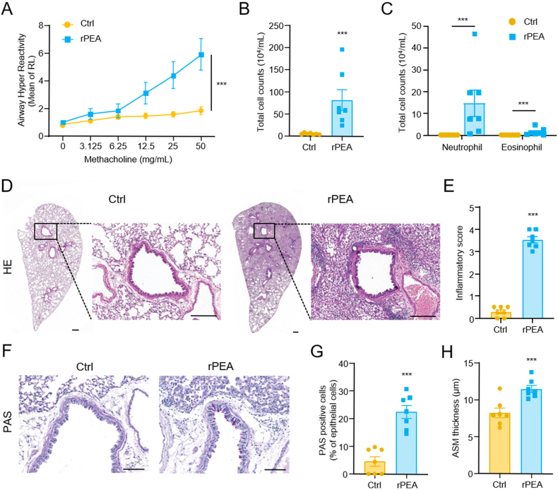
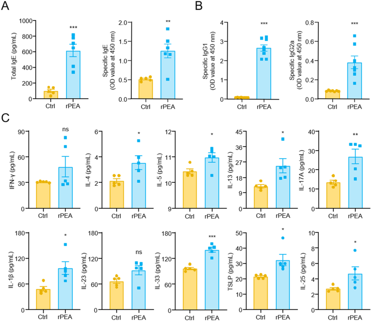
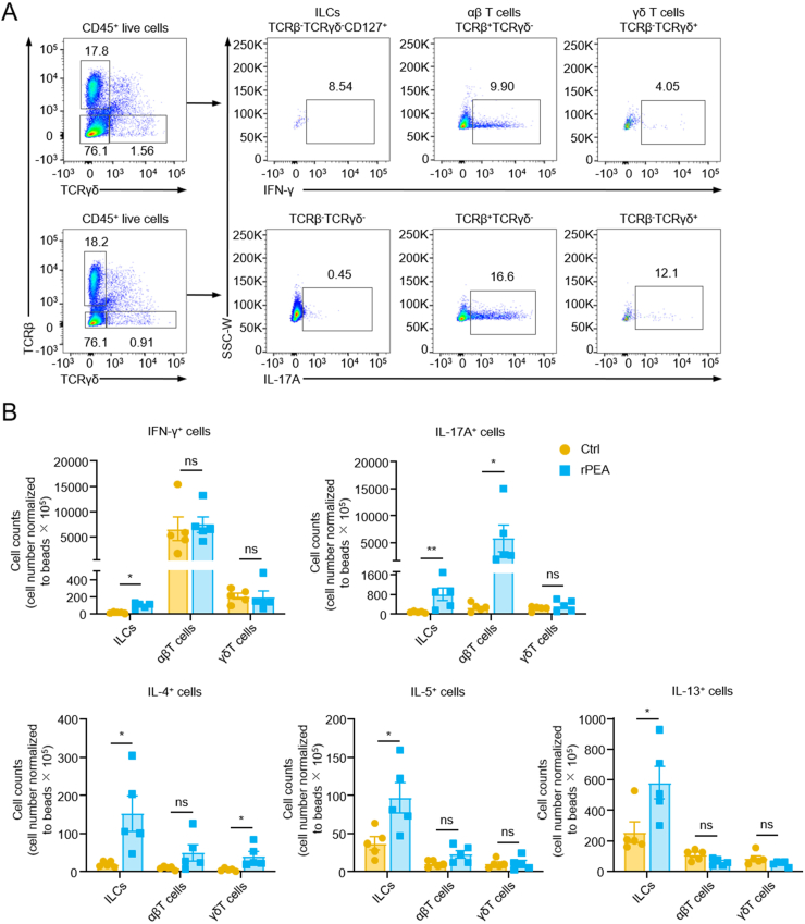
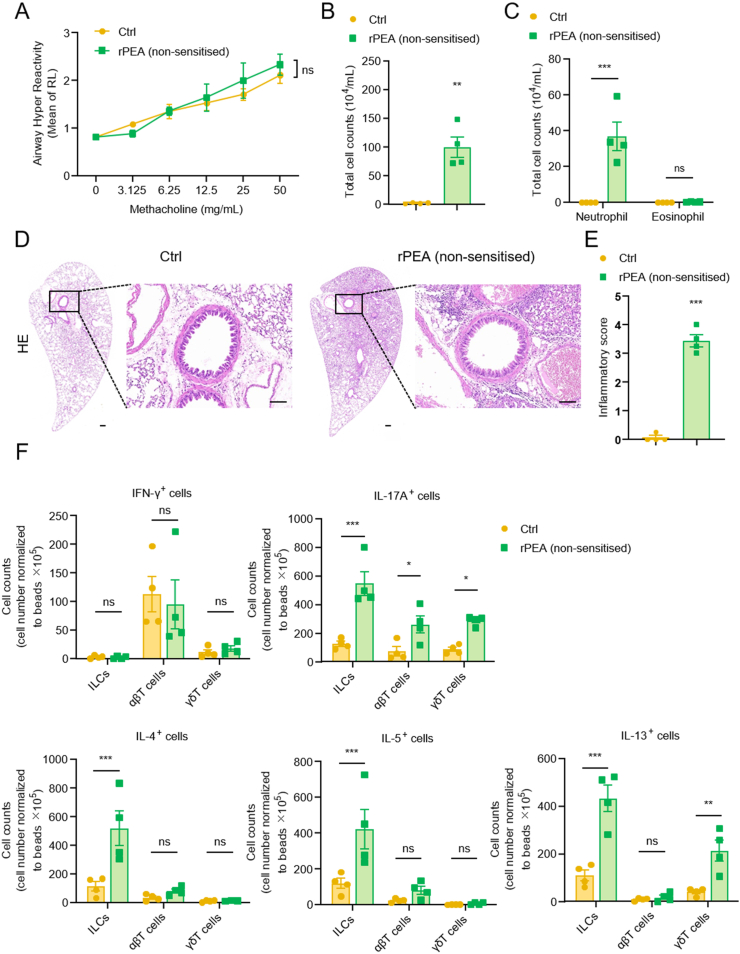
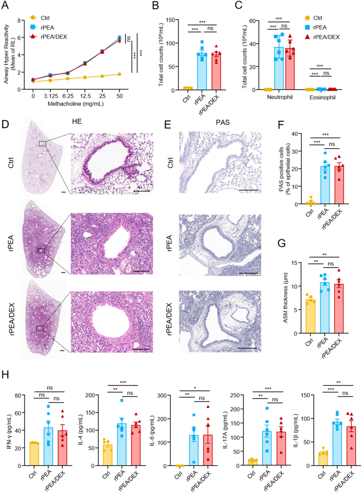
Results: PEA-specific IgE was identified in 17 (14.2 %) of 120 asthma patients. The rPEA-sensitized and challenged mice had increased PEA-specific immunoglobulins (such as IgE, IgG1 and IgG2a) and developed asthma-like phenotypes with airway hyperresponsiveness, severe airway inflammation, and airway remodeling. Lungs from these mice displayed significant increases in neutrophils and IL-17A+ cells. Innate lymphoid cells (ILCs) produced type 2 cytokines (IL-4, IL-5, and IL-13), whereas Th cells did not. Nonetheless, airway inflammation, rather than hyperresponsiveness, was elicited in non-sensitized mice upon challenge with rPEA. Importantly, rPEA-induced asthmatic mice were unresponsive to dexamethasone treatment.

Conclusion: PEA is a novel allergen that sensitizes asthmatic patients. Furthermore, mice developed steroid-resistant asthma, characterized by an atypical cytokine profile associated with non-TH2 inflammation, only after being sensitized and challenged with rPEA. These findings suggest a potentially significant role for PEA in asthma development, warranting consideration in clinical diagnosis and treatment strategies.

Keywords: Allergen; Asthma; IL-17A; Pseudomonas aeruginosa exotoxin A; Steroid-insensitive.


Conflict of interest statement

The authors declare that they have no known competing financial interests or personal relationships that could have appeared to influence the work reported in this paper.

Figures

Fig. 1
Fig. 1
Prokaryotic expression of rPEA and its serological identification in asthma patients. (A) The purity and apparent molecular weight of rPEA were determined by Coomassie brilliant blue. The target protein bands are located between 40 and 55 kDa. (B) Pie chart showing the proportion of positive and negative rPEA-sIgE in 120 asthma patients' sera. (C) Immunoblotting of IgE binding to rPEA using sera from six rPEA-sIgE positive patients (lanes 1–6). Samples of 2.5 μg per lane of rPEA was separated by electrophoresis on 12 % SDS-PAGE prior to transfer to PVDF membrane. Blots were incubated in patient sera (1:20) and then incubated with HRP-conjugated mouse anti-human IgE antibodies and visualized with ECL. M: molecular weight marker. BC: Blank control. NC: Negative control. (D) and (E) Specific IgE competitive ELISA assay. The rPEA-sIgE positive serum was incubated with increasing doses of rPEA (D) or P. aeruginosa lysate (E). The figure is shown as the inhibition of sIgE binding to plate-bound rPEA. The full, non-adjusted images of gels and blots are presented in Fig. S7.
Fig. 2
Fig. 2
Asthma-like pathological characteristics of rPEA-sensitized and challenged mice. (A) The airway hyperresponsiveness of mice was measured by lung resistance (RL) (n = 4–5). (B) Numbers of total inflammatory cell in bronchoalveolar lavage fluid (BALF) (n = 7). (C) Numbers of neutrophils and eosinophils in BALF (n = 7). (D) Representative HE-stained lung sections of mice. Scale bar: 200 μm. (E) Score of airway inflammation (n = 7). (F) Periodic acid-Schiff (PAS) stained lung sections of mice. Scale bar: 100 μm. (G) Quantification of PAS staining. The percentage of PAS positive cells in epithelial cells was calculated (n = 7). (H) Analysis of airway smooth muscle (ASM) thickness (n = 7). ***p < 0.001, Bar, mean ± SEM.
Fig. 3
Fig. 3
Levels of serum immunoglobulin E and BALF cytokines from rPEA-sensitized and challenged mice. (A) The total IgE and rPEA-specific IgE in sera (n = 5–6). The sera were diluted with 1:10. (B) Detection of rPEA specific IgG1 and IgG2a in sera (n = 7). The serum was diluted with 1:105. (C) Levels of IFN-γ, IL-4, IL-5, IL-13, IL-17A, IL-1β, IL-23, IL-33, TSLP, and IL-25 in BALF (n = 5). ns, not significant, *p < 0.05, **p < 0.01, ***p < 0.001. Bar, mean ± SEM.
Fig. 4
Fig. 4
A single-cell suspension of lung tissue from mice that had been sensitized and challenged with rPEA was investigated by flow cytometry. (A) Flow cytometry gating strategy for representative cytokines (IFN-γ and IL-17A) of innate lymphoid cells (ILCs), αβ T cells, and γδ T cells in the lung tissue. It is worth noting that NK cells need to be excluded with CD127 before evaluating IFN-γ of ILCs. The gating strategy of other cytokines (IL-4, IL-5 and IL-13) was the same as that of IL-17A (seen Fig. S2B). (B) Absolute number of IFN-γ, IL-4, IL-5, IL-13, IL-17A positive cells from ILCs, αβ T and γδ T cells (n = 5). ns, not significant, *p < 0.05, **p < 0.01. Bar, mean ± SEM.
Fig. 5
Fig. 5
Measures of airway hyperresponsiveness and lung inflammation in non-sensitized mice challenged with rPEA. (A) The airway hyperresponsiveness of mice was measured by lung resistance (RL) (n = 3–4). (B) Numbers of total inflammatory cell in bronchoalveolar lavage fluid (BALF) (n = 4). (C) Numbers of neutrophils and eosinophils in BALF (n = 4). (D) Representative HE-stained lung sections of mice. Scale bar: 200 μm. (E) Score of airway inflammation (n = 4). (F) Absolute number of IFN-γ, IL-4, IL-5, IL-13, IL-17A positive cells from ILCs, αβ T and γδ T cells (n = 5). ns, not significant, *p < 0.05, **p < 0.01, ***p < 0.001. Bar, mean ± SEM.
Fig. 6
Fig. 6
Asthma in mice induced by rPEA exposure are corticosteroid resistant. Dexamethasone (1 mg/kg) or PBS was daily administered i.p. 1 h before rPEA challenge on days 21–23. (A) The AHR of mice was measured by lung resistance (RL) (n = 5–6). (B) Numbers of total inflammatory cell in bronchoalveolar lavage fluid (BALF) (n = 5–6). (C) Differential BALF cell counts of neutrophils and eosinophils (n = 5–6). (D) Representative HE-stained lung sections of mice. Scale bar: 200 μm. (E) PAS-stained lung sections of mice. Scale bar: 200 μm. (F) Quantification of PAS staining. The percentage of PAS positive cells in epithelial cells was calculated (n = 5–6). (G) Analysis of airway smooth muscle (ASM) thickness (n = 5–6). (H) Levels of IFN-γ, IL-4, IL-6, IL-17A and IL-1β in BALF (n = 5–6). ns, not significant, *p < 0.05, **p < 0.01, ***p < 0.001. Bar, mean ± SEM.

Similar articles

References

    1. Hinks T.S.C., Levine S.J., Brusselle G.G. Treatment options in type-2 low asthma. Eur. Respir. J. 2021;57 doi: 10.1183/13993003.00528-2020. - DOI - PMC - PubMed
    1. Hammad H., Lambrecht B.N. The basic immunology of asthma. Cell. 2021;184:1469–1485. doi: 10.1016/j.cell.2021.02.016. - DOI - PubMed
    1. Barnes P.J. Targeting cytokines to treat asthma and chronic obstructive pulmonary disease. Nat. Rev. Immunol. 2018;18:454–466. doi: 10.1038/s41577-018-0006-6. - DOI - PubMed
    1. Lambrecht B., Hammad H. The immunology of asthma. Nat. Immunol. 2015;16:45–56. doi: 10.1038/ni.3049. - DOI - PubMed
    1. Kuwabara K., Hirose M., Kato K., Yokoi T., Shiga M., Kondo R., et al. Serological analysis of sensitization in allergic bronchopulmonary aspergillosis: a study on allergen components and interspecies relationships. J. Asthma. 2020;57:610–617. doi: 10.1080/02770903.2019.1599387. - DOI - PubMed

LinkOut - more resources