Case Reports
. 2024 Dec 20;40(1):48-59.
doi: 10.1093/ndt/gfae197.

Multidisciplinary team approach for CKD-associated osteoporosis

Affiliations
Case Reports

Multidisciplinary team approach for CKD-associated osteoporosis

Ditte Hansen et al. Nephrol Dial Transplant. .

Abstract

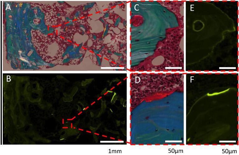
Chronic kidney disease-mineral and bone disorder (CKD-MBD) contributes substantially to the burden of cardiovascular disease and fractures in patients with CKD. An increasing arsenal of diagnostic tools, including bone turnover markers and bone imaging, is available to support clinicians in the management of CKD-associated osteoporosis. Although not mandatory, a bone biopsy remains useful in the diagnostic workup of complex cases. In this special report, the European Renal Osteodystrophy (EUROD) initiative introduces the concept of a kidney-bone multidisciplinary team (MDT) for the diagnosis and clinical management of challenging cases of CKD-associated osteoporosis. In 2021, the EUROD initiative launched virtual clinical-pathological case conferences to discuss challenging cases of patients with CKD-associated osteoporosis, in whom a bone biopsy was useful in the diagnostic workup. Out of these, we selected four representative cases and asked a kidney-bone MDT consisting of a nephrologist, an endocrinologist and a rheumatologist to provide comments on the diagnostic and therapeutic choices. These cases covered a broad spectrum of CKD-associated osteoporosis, including bone fracture in CKD G5D, post-transplant bone disease, disturbed bone mineralization, severely suppressed bone turnover and severe hyperparathyroidism. Comments from the MDT were, in most cases, complementary to each other and additive to the presented approach in the cases. The MDT approach may thus set the stage for improved diagnostics and tailored therapies in the field of CKD-associated osteoporosis. We demonstrate the clinical utility of a kidney-bone MDT for the management of patients with CKD-MBD and recommend their establishment at local, national, and international levels.

Keywords: bone biopsy; chronic kidney disease–mineral and bone disorder; multidisciplinary team; osteoporosis; renal osteodystrophy.


Conflict of interest statement

D.H. reports research grant from Vifor Pharma and Gedeon Richter and consultancy fees and lecture fees from UCB Nordic, GSK and AstraZeneca. A.C.F. reports lecture fees from Vifor and AstraZeneca. R.J. received fees as site PI in a pharmacy-initiated study (SHP634-401) of Takeda. S.K. reports consultancy fees from Vifor Pharma, GSK and Bayer, and lecture fees from AstraZeneca. M.H.L.-P. reports research grant from Kiowa Kirin. K.E.S.P. educational fora lecture fees and honoraria from UCB and Amgen, with all fees donated to charity via waiver before undertaking the work. P.E. reports research grant from Vifor Pharma, and consultancy fees and lecture fees from UCB and Vifor Pharma. M.H. advisory board Resverlogix and employee at Diaverum AB. H.S.J., T.L.A., A.F., H.K., L.M., and X.T. report no conflicts of interest.

Figures

Graphical Abstract
Graphical Abstract
Figure 1:
Figure 1:
Case 1: bone biopsy from a patient with CKD G4 and a history of sarcoidosis presenting with low BMD and high bone-specific alkaline phosphatase. The biopsy revealed low trabecular bone volume with increased cortical porosity. Bone turnover was normal to high, with increased bone resorption. There was no mineralization defect. Full section of iliac crest sample. Goldner-stained section (5×). Asterisks illustrate the increase in cortical porosity of the outer cortex. Dotted line marks the space where the inner cortex was before being trabecularized and resorbed. Short arrows illustrate the extreme thinning of the remaining inner cortex.
Figure 2:
Figure 2:
Trends of mineral metabolism parameters and bone biopsy—Case 2: a patient with a kidney transplant presenting with hypercalcemic hyperparathyroidism. (A) Trends of mineral metabolism parameters. Ca: calcium. (B) A bone biopsy was performed at times 0 and at 24 months. The bone biopsy at 24 months revealed high turnover bone disease. Active bone resorption was increased, while osteoid parameters were in the normal range. UV microscopy showed abundant double labels. TMV classification: turnover: high; mineralization: normal; volume: low. (B1, B2) Masson Goldner's trichrome stain; (B3, B4) fluorescent labelling. Scale bars: 2 mm (B1, B3); 200 µm (B2, B4).
Figure 3:
Figure 3:
Trends of mineral metabolism parameters and bone biopsy—Case 3: a patient with diabetes and kidney transplantation presenting with bone pain and multiple low-energy fracturs. (A) Trends of mineral metabolism parameters. Ca: calcium. (B) Bone biopsy showed low trabecular bone volume and thin cortical bone (A) with abnormal mineralization (B, C, E) and low turnover (B, D, F). (A, C, E) Partly polarized light digital microscopy of Masson trichrome staining, showing osteoid surfaces mainly directly deposited on eroded surfaces colonized by a low density of osteoblasts. (B, D, F) Fluorescence digital microscopy of tetracyclin-labeled bone surfaces, which only represented a small fraction of the osteoid surfaces. Scale bars: 1 mm (A, B); 50 µm (C–F).
Figure 4:
Figure 4:
Bone biopsy—Case 4: a patient on hemodialysis presenting with multiple low-energy fractures and low-normal PTH. Bone biopsy showed low trabecular bone volume (A) with normal turnover (B, E, F) and normal mineralization (C, D). Double labels occurred, but this was not included in the high magnification illustration. (A, C, D) Partly polarized light digital microscopy of Masson trichrome staining. (B, E, F) Fluorescence digital microscopy of tetracyclin-labeled bone surfaces. Scale bars: 1 mm (A, B); 50 µm (C–F).
Figure 5:
Figure 5:
Work flow kidney–bone MDTs.

References

    1. Aguilar A, Gifre L, Ureña-Torres Pet al. . Pathophysiology of bone disease in chronic kidney disease: from basics to renal osteodystrophy and osteoporosis. Front Physiol 2023;14:1177829. 10.3389/fphys.2023.1177829 - DOI - PMC - PubMed
    1. Magagnoli L, Cozzolino M, Caskey FJet al. . Association between CKD-MBD and mortality in older patients with advanced CKD—results from the EQUAL study. Nephrol Dial Transplant 2023;38:2562–75. 10.1093/ndt/gfad100 - DOI - PMC - PubMed
    1. Yamada S, Nakano T. Role of chronic kidney disease (CKD)-mineral and bone disorder (MBD) in the pathogenesis of cardiovascular disease in CKD. J Atheroscler Thromb 2023;30:835. 10.5551/jat.RV22006 - DOI - PMC - PubMed
    1. Hampson G, Elder GJ, Cohen-Solal Met al. . A review and perspective on the assessment, management and prevention of fragility fractures in patients with osteoporosis and chronic kidney disease. Endocrine 2021;73:509–29. 10.1007/s12020-021-02735-9 - DOI - PMC - PubMed
    1. Jørgensen HS, Evenepoel P. Persistent hyperparathyroidism: a reality calling for additional evidence. Am J Kidney Dis 2023;81:256–8. 10.1053/j.ajkd.2022.10.001 - DOI - PubMed

Publication types

MeSH terms