. 2024 Oct 16;15(10):e0181224.
doi: 10.1128/mbio.01812-24. Epub 2024 Sep 24.

Growth defect of domain III glycoprotein B mutants of human cytomegalovirus reverted by compensatory mutations co-localizing in post-fusion conformation

Affiliations

Growth defect of domain III glycoprotein B mutants of human cytomegalovirus reverted by compensatory mutations co-localizing in post-fusion conformation

Madlen Mollik et al. mBio. .

Abstract

Cell entry is a crucial step for a virus to infect a host cell. Human cytomegalovirus utilizes glycoprotein B (gB) to fuse the viral and host cell membranes upon receptor binding of gH/gL-containing complexes. Fusion is mediated by major conformational changes of gB from a metastable pre-fusion to a stable post-fusion state whereby the central trimeric coiled-coils, formed by domain (Dom)III α helices, remain structurally nearly unchanged. To better understand the role of the stable core, we individually introduced three potentially helix-breaking or one disulfide bond-breaking mutation in the DIII α3 to study different aspects of the viral behavior upon long-term culturing. Two of the three helix-breaking mutations, gB_Y494P and gB_I495P, were lethal for the virus in either fibroblasts or epithelial cells. The third substitution, gB_G493P, on the other hand, displayed a delayed replication and spread, which was more pronounced in epithelial cells, hinting at an impaired fusion. Interestingly, the disulfide bond-breaker mutation, gB_C507S, performed strikingly differently in the two cell types - lethal in epithelial cells and an atypical phenotype in fibroblasts, respectively. Replication curve analyses paired with the infection efficiency, the spread morphology, and the cell-cell fusogenicity suggest a dysregulated fusion process, which could be reverted by second-site mutations mapping predominantly to gB DomV. Our findings underline the functional importance of a stable DomIII core for a well-regulated DomV rearrangement during fusion.IMPORTANCEHuman cytomegalovirus (HCMV) can establish a lifelong infection. In most people, the infection follows an asymptomatic course; however, it is a major cause of morbidity and mortality in immunocompromised patients or neonates. HCMV has a very broad cell tropism, ranging from fibroblasts to epi- and endothelial cells. The virus uses different entry pathways utilizing the core fusion machinery consisting of glycoprotein complexes gH/gL and glycoprotein B (gB). The fusion protein gB undergoes fundamental rearrangements from a metastable pre-fusion to a stable post-fusion conformation. Here, we characterized the viral behavior after the introduction of four single-point mutations in the gB central core. These led to various cell type-specific atypical phenotypes and the emergence of compensatory mutations, demonstrating an important interaction between domains III and V. We provide a new basis for the development of a structurally and functionally altered gB, which can further serve as a tool for drug and vaccine development.

Keywords: entry and spread; glycoprotein B; glycoprotein O; human cytomegalovirus; membrane fusion.


Conflict of interest statement

The authors declare no conflict of interest.

Figures

Fig 1
Fig 1
Schematic of TB40-BAC4-luc DNA and protomer gB pre-fusion structure. (A) Illustration of TB40-BAC4-luc DNA with main genome characteristics used for the transfection and reconstitution in fibroblasts and epithelial cells. Diagram of gB encoded by UL55 displaying the C111-C507 disulfide bond; fusion loops are indicated with a purple line, the furin cleavage site is marked with a dotted black line and the five structural gB DomI–V, MPR, and TM in color. Enlarged the sequence alignment of the α3 helix with representative sequences of HCMV, HSV-1, VZV, and PrV as indicated. Location of the mutations are depicted by dotted black boxes in the alignment. (B) Protomer structure (PDB: 7KDP). Mutation sites G493-I495 and C507, forming a disulfide bond with C111, are marked in black. gB domains are colored as follows: DomI (dark blue), DomII (green), DomIII (yellow), DomIV (orange), DomV (red), the membrane proximal region (MPR, blue), and the transmembrane domain (TM, turquoise) created with UCSF ChimeraX [61]. GT, genotype; HSV-1, herpes simplex virus 1; VZV, varicella zoster virus; PrV, pseudorabies virus; N-term, N-terminal region; Cyto, cytoplasmic tail.
Fig 2
Fig 2
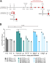
Viral replication and infectivity differences among gB mutants independent of gO genotypes. (A and B) HFF or ARPE-19 were individually transfected in duplicates with the respective TB40-BAC-luc DNA and long-term cultured. During reconstitution, cell-free samples were taken from the supernatant once a week during medium change, and cell-associated samples were taken during passaging (P1–P3) as indicated. (A and B) Top panel: viral DNA released into the supernatant of transfected HFF (A) or ARPE-19 cells (B) with the parental gB_TB40gOGT1c in black, gB_G493P mutants in purple and red, and gB_C507S mutants in petrol and light blue, each in gOGT1c and gOGT1c3 background, respectively. (A and B) Bottom panel: viral DNA load in cell-associated samples of transfected HFF (A) or ARPE-19 cells (B) normalized to the genomic DNA. (C) Subsequent to the harvest of all gB clones, the infection efficiency of cell-free viruses was tested in parallel through a luciferase assay. After infection, HFFs were incubated for 48 h and ARPE-19 cells for 72 h before the determination of relative light units (RLUs) in the cell lysates as the read-out of infection efficiency. Positive samples on HFFs were then tested on ARPE-19 cells (right). P, passaging; gDNA, genomic DNA.
Fig 3
Fig 3
Increase in cell-free infectivity of gB_C507S mutant viral particles late after transfection and further propagation. (A) Experimental scheme showing the transfection and infection period with the time points indicating the main characteristics: passaging (P1 and P2) and pre- and post-phenotype change of C507S mutants in gray dots and initial and further release of infectious virus into supernatant (cell-free) until harvest post-transfection and post-infection in red dots. (B) Infectious cell-free parental and gB_C507S virus stocks at the indicated dpt and dpi were investigated for (i) the number of encapsidated genome particles and (ii) the infection efficiency. Encapsidated genome copy numbers were determined by HCMV-specific quantitative PCR after DNase I treatment. Infection capacity was assessed in parallel 48 h after HFF infection by measuring the RLUs in cell lysates. Calculated particle-to-RLU ratios in log10 are plotted for parental and gB_C507S mutant clones 1 (black and colored columns) and 2 (open columns) post-transfection and post-infection (columns with patterns). Colors for the mutant clones are the same as in Fig. 2. Columns indicate the mean. Error bars are the standard deviation. A comparison of the medians from two clones in each group (in duplicate) as indicated by the brackets was performed using the non-parametric Kruskal–Wallis test and Dunn’s post hoc test for multiple comparisons. ns, not statistically significant (P > 0.05).
Fig 4
Fig 4
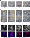
Morphology change of gB_C507S mutant foci on fibroblasts during long-term culturing. (A and B) After transfection of HFFs with the parental (A) and the gB mutant TB40-BAC-luc DNA clones (B) as indicated, light microscopy (Leica) pictures were regularly taken during long-term culturing indicated with dpt. Representative images (10×) of cytopathic effects of parental strain (A) compared to gB_C507S mutants (B), displaying highly dense foci at 51 dpt and beginning to change the phenotype to an evenly spread morphology at around 63 dpt. The black boxes highlight the areas shown on the right. White arrows indicate the definable (left panels) and the indistinct (right panels) borders of the foci to the surrounding cell monolayer. (C) gB_C507S mutants harvested prior to (left panels) and after (right panels) the phenotype change were subjected to immediate early (IE) and DAPI staining 22 days after HFF infection (dpi) when the characteristic phenotype was present. The white boxes highlight the areas shown on the right. Representative plaques of the focal growth and the even spread are shown in 10×. (1), clone 1; (2), clone 2; PH, phase contrast; FITC, fluorescein isothiocyanate; DAPI, 4′,6-diamidino-2-phenylindole.
Fig 5
Fig 5
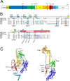
Co-localization of second-site mutations of gB_C507S mutants in gB post-fusion structure. (A) Schematic diagram of linear gB marked with second-site mutations as black lines that emerged in all gB_C507S clones during the post-transfection and post-infection period (see Table 2). (B) Amino acid alignments of gB from two HCMV strains (TB40 and Towne) and three alphaherpesviruses (HSV-1, VZV, and PrV) as indicated with the homologous sites of the second-site mutations highlighted with black boxes. Respective substitutions are shown below. Broken circles indicate the residues in HSV-1 and PrV for which fusion mutants have been described (41–43). (C) Protomer structure of gB with C507 in α3 helix marked in black. Acquired second-site mutations are highlighted in pre-fusion (left; PDB: 7KDP) and post-fusion protomer (right; PDB: 5C6T) with the majority of those located in the encircled post-fusion region. Created with UCSF ChimeraX [61]. Domains in all depictions are colored as in Fig. 1. α, alpha helix; β, beta sheet.
Fig 6
Fig 6
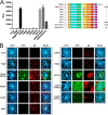
Compensation of the cell–cell fusion defect of gB_C507S by second-site mutations in gB domain V. (A) The 293T-DSP-mix cells were transiently transfected with an empty vector (mock), wild-type full-length TB40gB (TBgB), the intrinsically fusion-competent TBgB/VSV-G without mutations, or any of the indicated mutated versions thereof. Cell–cell fusions were quantified via bioluminescence, given in RLUs depicted as mean values of biological triplicates ± standard deviations. (B) Immunofluorescence analyses of 293T-DSP-mix cells were performed 3 days after transfection with the same expression plasmids as used in (A). After fixation, cell nuclei were visualized using DAPI and the GFP signal of the reconstituted DSP-reporter protein upon cell–cell fusion. As a control, gB expression was determined by anti-gB MAb 27-287. Representative confocal laser scanning microscopy images are given. TBgB/VSV-G, chimeric version of gB (aa 1–750 of strain TB40E; ABV71586.1) with vesicular stomatitis virus G protein (VSV-G) transmembrane and cytoplasmic tail as previously described (44); mock, empty pcDNA3 vector; MAb, monoclonal antibody.

References

    1. Ebel H, Benecke T, Vollmer B. 2022. Stabilisation of viral membrane fusion proteins in prefusion conformation by structure-based design for structure determination and vaccine development. Viruses 14:1816. doi:10.3390/v14081816 - DOI - PMC - PubMed
    1. Connolly SA, Jardetzky TS, Longnecker R. 2021. The structural basis of herpesvirus entry. Nat Rev Microbiol 19:110–121. doi:10.1038/s41579-020-00448-w - DOI - PMC - PubMed
    1. Lopper M, Compton T. 2002. Disulfide bond configuration of human cytomegalovirus glycoprotein B. J Virol 76:6073–6082. doi:10.1128/jvi.76.12.6073-6082.2002 - DOI - PMC - PubMed
    1. Stangherlin LM, de Paula FN, Icimoto MY, Ruiz LGP, Nogueira ML, Braz ASK, Juliano L, da Silva MCC. 2017. Positively selected sites at HCMV gB furin processing region and their effects in cleavage efficiency. Front Microbiol 8:934. doi:10.3389/fmicb.2017.00934 - DOI - PMC - PubMed
    1. Burke HG, Heldwein EE. 2015. Crystal structure of the human cytomegalovirus glycoprotein B. PLoS Pathog 11:e1005227. doi:10.1371/journal.ppat.1005227 - DOI - PMC - PubMed

Substances

LinkOut - more resources