. 2024 Oct 7;221(10):e20240637.
doi: 10.1084/jem.20240637. Epub 2024 Sep 24.

Autoantibodies neutralizing type I IFNs underlie severe tick-borne encephalitis in ∼10% of patients

Affiliations

Autoantibodies neutralizing type I IFNs underlie severe tick-borne encephalitis in ∼10% of patients

Adrian Gervais et al. J Exp Med. .

Abstract

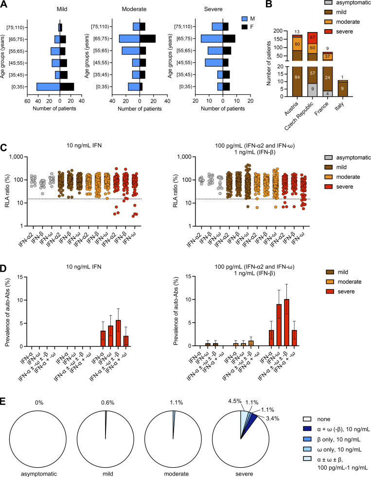
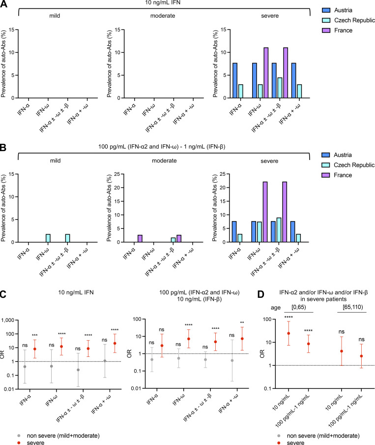
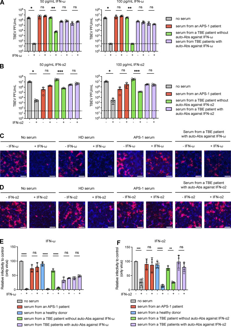
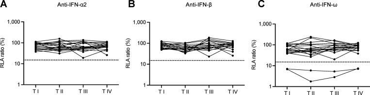
Tick-borne encephalitis (TBE) virus (TBEV) is transmitted to humans via tick bites. Infection is benign in >90% of the cases but can cause mild (<5%), moderate (<4%), or severe (<1%) encephalitis. We show here that ∼10% of patients hospitalized for severe TBE in cohorts from Austria, Czech Republic, and France carry auto-Abs neutralizing IFN-α2, -β, and/or -ω at the onset of disease, contrasting with only ∼1% of patients with moderate and mild TBE. These auto-Abs were found in two of eight patients who died and none of 13 with silent infection. The odds ratios (OR) for severe TBE in individuals with these auto-Abs relative to those without them in the general population were 4.9 (95% CI: 1.5-15.9, P < 0.0001) for the neutralization of only 100 pg/ml IFN-α2 and/or -ω, and 20.8 (95% CI: 4.5-97.4, P < 0.0001) for the neutralization of 10 ng/ml IFN-α2 and -ω. Auto-Abs neutralizing type I IFNs accounted for ∼10% of severe TBE cases in these three European cohorts.


Conflict of interest statement

Disclosures: The Center for Virology, Medical University of Vienna, Austria, received an Investigator-Initiated Research grant from Pfizer (WI235042), with J.H. Aberle as principal investigator, which is not related to the submitted work. J.-L. Casanova reported a patent to PCT/US2021/042741 pending. No other disclosures were reported.

Figures

Figure 1.
Figure 1.
Demographic and phenotypic distributions of the TBE cohort and auto-Abs neutralizing type I IFNs in individuals infected with TBEV. (A) Age and sex distribution of the patients according to TBE severity. (B) Composition of the TBE cohort: number of individuals in each TBE group enrolled at each center. (C) Luciferase-based neutralization assay to detect auto-Abs neutralizing 10 ng/ml IFN-α2, IFN-ω, or IFN-β (left panel) and 100 pg/ml IFN-α2 or IFN-ω, or 1 ng/ml IFN-β (right panel). Plasma samples from asymptomatic TBE cases (gray), patients with mild TBE (brown), patients with moderate TBE (orange), and patients with severe TBE (red) were diluted 1:10. HEK293T cells were transfected with (1) a plasmid containing the firefly luciferase gene under the control of an ISRE-containing promotor and (2) a plasmid containing the Renilla luciferase gene. The cells were then treated with type I IFNs and RLA was calculated by normalizing firefly luciferase activity against Renilla luciferase activity. An RLA <15% of the median RLA for healthy controls was considered to correspond to neutralizing activity (dotted line; Bastard et al., 2021a). Each sample was tested once. (D) Proportions of individuals with auto-Abs neutralizing type I IFNs at a concentration of 10 ng/ml (left) or 1 ng/ml–100 pg/ml (right) in the three groups of TBE patients (mild, moderate, severe), as determined with the luciferase-based neutralization assay. IFN-α, auto-Abs neutralizing IFN-α2 (regardless of their effects on other IFNs); IFN-ω, auto-Abs neutralizing IFN-ω (regardless of their effects on other IFNs); IFN-α ± ω ± β, auto-Abs neutralizing IFN-α2 and/or IFN-ω and/or IFN-β; IFN-α + ω, auto-Abs neutralizing both IFN-α2 and IFN-ω. The bars indicate the upper and lower limits of the 95% CI. (E) Proportion of type I IFNs neutralized in the three groups of TBE patients (mild, moderate, severe) and in individuals with silent infection according to the nature and combination of auto-Abs.
Figure S1.
Figure S1.
Correlation between ELISA and neutralization assay results for the detection of auto-Abs neutralizing type I IFNs.
Figure 2.
Figure 2.
Comparison of the proportions of individuals positive for auto-Abs neutralizing type I IFNs in the different TBE cohorts and estimation of the risk of severe TBE in auto-Ab-positive individuals relative to the general population. (A) Comparison of auto-Ab prevalence in TBE patients from the Austrian, Czech, and French cohorts (considering auto-Abs neutralizing 10 ng/ml IFN). IFN-α, auto-Abs neutralizing IFN-α2 (regardless of their effects on other IFNs); IFN-ω, auto-Abs neutralizing IFN-ω (regardless of their effects on other IFNs); IFN-α ± ω ± β, auto-Abs neutralizing IFN-α2 and/or IFN-ω and/or IFN-β; IFN-α + ω, auto-Abs neutralizing both IFN-α2 and IFN-ω. (B) Comparison of auto-Ab prevalence in TBE patients from the Austrian, Czech, and French cohorts (considering auto-Abs neutralizing 100 pg/ml [IFN-α2 and IFN-ω] or 1 ng/ml [IFN-β]). IFN-α, auto-Abs neutralizing IFN-α2 (regardless of their effects on other IFNs); IFN-ω, auto-Abs neutralizing IFN-ω (regardless of their effects on other IFNs); IFN-α ± ω ± β, auto-Abs neutralizing IFN-α2 and/or IFN-ω and/or IFN-β; IFN-α + ω, auto-Abs neutralizing both IFN-α2 and IFN-ω. (C) OR for the presence of auto-Abs in individuals with non-severe (gray) or severe (red) TBE relative to the general population, with adjustment for age and sex by logistic regression. The horizontal bars indicate the upper and lower limits of the 95% CI. IFN-α, auto-Abs neutralizing IFN-α2 (regardless of their effects on other IFNs); IFN-ω, auto-Abs neutralizing IFN-ω (regardless of their effects on other IFNs); IFN-α ± ω ± β, auto-Abs neutralizing IFN-α2 and/or IFN-ω and/or IFN-β; IFN-α + ω, auto-Abs neutralizing both IFN-α2 and IFN-ω. (D) OR for the presence of auto-Abs in individuals with severe TBE relative to the general population by age group, with adjustment for sex by logistic regression. ORs were calculated separately for patients with severe TBE aged ≤65 and >65 years. The horizontal bars indicate the upper and lower limits of the 95% CI. IFN-α, auto-Abs neutralizing IFN-α2 (regardless of their effects on other IFNs); IFN-ω, auto-Abs neutralizing IFN-ω (regardless of their effects on other IFNs); IFN-α ± ω ± β, auto-Abs neutralizing IFN-α2 and/or IFN-ω and/or IFN-β; IFN-α + ω, auto-Abs neutralizing both IFN-α2 and IFN-ω. [0,65): individuals aged 0–65 years old; [65,110): individuals aged 65–110 years old; ns: non-significant; **P < 0.01, ***P < 0.001, ****P < 0.0001.
Figure S2.
Figure S2.
Anti-TBEV IgG and IgM titers in the Austrian and French cohorts of TBE patients, by disease severity. (A and B) The blue arrows show the patients with auto-Abs neutralizing type I IFNs. The dotted lines represent the thresholds for positivity. The assays used to detect anti-TBEV IgG and IgM titers differed between the two cohorts, accounting for the difference in positivity thresholds.
Figure 3.
Figure 3.
TBEV infection and IFN treatment in Vero-E6 cells. (A and B) Plaque assay showing TBEV viral titers (PFU/ml) in Vero-E6 cells left untreated or treated with serum samples from patients with or without auto-Abs neutralizing IFN-ω in the presence of 50 pg/ml IFN-ω (A, left panel), 100 pg/ml IFN-ω (A, right panel), 50 pg/ml IFN-α2 (B, left panel), or 100 pg/ml IFN-α2 (B, right panel). For each set of serum conditions, we compared the values obtained in the presence and absence of IFN treatment in an ordinary one-way ANOVA with Bonferroni correction (as implemented in GraphPad Prism version 10.2.3). ns: non-significant; *: P < 0.05; **: P < 0.01; ***: P < 0.001. The dashed line indicates the limit of detection of the plaque assay. (C and D) Fluorescence microscopy images of Vero-E6 cells with and without treatment with 50 pg/ml IFN-ω (C) or 50 pg/ml IFN-α2 (D) in the absence of serum or the presence of serum from a healthy donor (HD), an APS-1 patient (with auto-Abs against IFN-ω and IFN-α2), a TBE patient with auto-Abs against IFN-ω (C) or a TBE patient with auto-Abs against IFN-α2 (D). Vero-E6 cells were then infected with mCherry-TBEV. The nuclei were stained with Hoechst stain before the measurement of fluorescence. The scale bars represent 300 µm. (E and F) Quantification of TBEV-positive Vero-E6 cells normalized against the percentage of cells infected in TBEV-only conditions, for different serum samples, with or without IFN-ω (E) or IFN-α2 (F) treatment. ns: non-significant; **: P < 0.01; ***: P < 0.001; ****: P < 0.0001. For each set of serum conditions, the values obtained in the presence and absence of IFN treatment were compared in an ordinary one-way ANOVA with Bonferroni correction (as implemented in GraphPad Prism version 10.2.3).
Figure 4.
Figure 4.
Auto-Abs neutralizing type I IFNs in longitudinal samples. (A–C) Auto-Abs neutralizing IFN-α2 (A), IFN-ω (B), and IFN-β (C), as determined with the luciferase-based neutralization assay, at different time points after infection. Each sample was tested once with an IFN concentration of 100 pg/ml (IFN-α2 and IFN-ω) or 1 ng/ml (IFN-β). I: time of admission to hospital, II: 2 days after admission, III: hospital discharge, and IV: follow-up (1–3 mo).

Similar articles

  • Auto-Abs neutralizing type I IFNs in patients with severe Powassan, Usutu, or Ross River virus disease.
    Gervais A, Bastard P, Bizien L, Delifer C, Tiberghien P, Rodrigo C, Trespidi F, Angelini M, Rossini G, Lazzarotto T, Conti F, Cassaniti I, Baldanti F, Rovida F, Ferrari A, Mileto D, Mancon A, Abel L, Puel A, Cobat A, Rice CM, Cadar D, Schmidt-Chanasit J, Scheid JF, Lemieux JE, Rosenberg ES, Agudelo M, Tangye SG, Borghesi A, Durand GA, Duburcq-Gury E, Valencia BM, Lloyd AR, Nagy A, MacDonald MM, Simonin Y, Zhang SY, Casanova JL. Gervais A, et al. J Exp Med. 2024 Dec 2;221(12):e20240942. doi: 10.1084/jem.20240942. Epub 2024 Nov 1. J Exp Med. 2024. PMID: 39485284 Free PMC article.
  • Autoantibodies neutralizing type I IFNs underlie West Nile virus encephalitis in ∼40% of patients.
    Gervais A, Rovida F, Avanzini MA, Croce S, Marchal A, Lin SC, Ferrari A, Thorball CW, Constant O, Le Voyer T, Philippot Q, Rosain J, Angelini M, Pérez Lorenzo M, Bizien L, Achille C, Trespidi F, Burdino E, Cassaniti I, Lilleri D, Fornara C, Sammartino JC, Cereda D, Marrocu C, Piralla A, Valsecchi C, Ricagno S, Cogo P, Neth O, Marín-Cruz I, Pacenti M, Sinigaglia A, Trevisan M, Volpe A, Marzollo A, Conti F, Lazzarotto T, Pession A, Viale P, Fellay J, Ghirardello S, Aubart M, Ghisetti V, Aiuti A, Jouanguy E, Bastard P, Percivalle E, Baldanti F, Puel A, MacDonald MR, Rice CM, Rossini G, Murray KO, Simonin Y, Nagy A, Barzon L, Abel L, Diamond MS, Cobat A, Zhang SY, Casanova JL, Borghesi A. Gervais A, et al. J Exp Med. 2023 Sep 4;220(9):e20230661. doi: 10.1084/jem.20230661. Epub 2023 Jun 22. J Exp Med. 2023. PMID: 37347462 Free PMC article.
  • Tick-borne encephalitis among U.S. travelers to Europe and Asia - 2000-2009.
    Centers for Disease Control and Prevention (CDC). Centers for Disease Control and Prevention (CDC). MMWR Morb Mortal Wkly Rep. 2010 Mar 26;59(11):335-8. MMWR Morb Mortal Wkly Rep. 2010. PMID: 20339345
  • Tick-borne encephalitis in Europe and Russia: Review of pathogenesis, clinical features, therapy, and vaccines.
    Ruzek D, Avšič Županc T, Borde J, Chrdle A, Eyer L, Karganova G, Kholodilov I, Knap N, Kozlovskaya L, Matveev A, Miller AD, Osolodkin DI, Överby AK, Tikunova N, Tkachev S, Zajkowska J. Ruzek D, et al. Antiviral Res. 2019 Apr;164:23-51. doi: 10.1016/j.antiviral.2019.01.014. Epub 2019 Jan 31. Antiviral Res. 2019. PMID: 30710567 Review.
  • A newly detected tick-borne encephalitis (TBE) focus in south-east Sweden: a follow-up study of TBE virus (TBEV) seroprevalence.
    Stjernberg L, Holmkvist K, Berglund J. Stjernberg L, et al. Scand J Infect Dis. 2008;40(1):4-10. doi: 10.1080/00365540701522934. Epub 2007 Sep 12. Scand J Infect Dis. 2008. PMID: 17852896 Review.

Cited by

References

    1. Ackermann-Gäumann, R., Eyer C., Vock M., Gowland P., Tinguely C., Leib S.L., Bori M., Buser A., Fontana S., Thierbach J., et al. . 2023. Prevalence of anti-tick-borne encephalitis virus (TBEV) antibodies in Swiss blood donors in 2014-2015. Blood Transfus. 21:100–109. 10.2450/2022.0099-22 - DOI - PMC - PubMed
    1. Agudelo, M., Palus M., Keeffe J.R., Bianchini F., Svoboda P., Salát J., Peace A., Gazumyan A., Cipolla M., Kapoor T., et al. . 2021. Broad and potent neutralizing human antibodies to tick-borne flaviviruses protect mice from disease. J. Exp. Med. 218:e20210236. 10.1084/jem.20210236 - DOI - PMC - PubMed
    1. Alotaibi, F., Alharbi N.K., Rosen L.B., Asiri A.Y., Assiri A.M., Balkhy H.H., Al Jeraisy M., Mandourah Y., AlJohani S., Al Harbi S., et al. . 2023. Type I interferon autoantibodies in hospitalized patients with Middle East respiratory syndrome and association with outcomes and treatment effect of interferon beta-1b in MIRACLE clinical trial. Influenza Other Respir. Viruses. 17:e13116. 10.1111/irv.13116 - DOI - PMC - PubMed
    1. Bastard, P., Gervais A., Le Voyer T., Philippot Q., Cobat A., Rosain J., Jouanguy E., Abel L., Zhang S.-Y., Zhang Q., et al. . 2024a. Human autoantibodies neutralizing type I IFNs: From 1981 to 2023. Immunol. Rev. 322:98–112. 10.1111/imr.13304 - DOI - PMC - PubMed
    1. Bastard, P., Gervais A., Le Voyer T., Rosain J., Philippot Q., Manry J., Michailidis E., Hoffmann H.H., Eto S., Garcia-Prat M., et al. . 2021a. Autoantibodies neutralizing type I IFNs are present in ∼4% of uninfected individuals over 70 years old and account for ∼20% of COVID-19 deaths. Sci. Immunol. 6:eabl4340. 10.1126/sciimmunol.abl4340 - DOI - PMC - PubMed

LinkOut - more resources