Structural basis for adhesin secretion by the outer-membrane usher in type 1 pili
- PMID: 39316053
- PMCID: PMC11459180
- DOI: 10.1073/pnas.2410594121
Structural basis for adhesin secretion by the outer-membrane usher in type 1 pili
Abstract
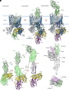
Gram-negative bacteria produce chaperone-usher pathway pili, which are extracellular protein fibers tipped with an adhesive protein that binds to a receptor with stereochemical specificity to determine host and tissue tropism. The outer-membrane usher protein, together with a periplasmic chaperone, assembles thousands of pilin subunits into a highly ordered pilus fiber. The tip adhesin in complex with its cognate chaperone activates the usher to allow extrusion across the outer membrane. The structural requirements to translocate the adhesin through the usher pore from the periplasm to the extracellular space remains incompletely understood. Here, we present a cryoelectron microscopy structure of a quaternary tip complex in the type 1 pilus system from Escherichia coli, which consists of the usher FimD, chaperone FimC, adhesin FimH, and the tip adapter FimF. In this structure, the usher FimD is caught in the act of secreting its cognate adhesin FimH. Comparison with previous structures depicting the adhesin either first entering or having completely exited the usher pore reveals remarkable structural plasticity of the two-domain adhesin during translocation. Moreover, a piliation assay demonstrated that the structural plasticity, enabled by a flexible linker between the two domains, is a prerequisite for adhesin translocation through the usher pore and thus pilus biogenesis. Overall, this study provides molecular details of adhesin translocation across the outer membrane and elucidates a unique conformational state adopted by the adhesin during stepwise secretion through the usher pore. This study elucidates fundamental aspects of FimH and usher dynamics critical in urinary tract infections and is leading to antibiotic-sparing therapeutics.
Keywords: adhsein FimH; outer-membrane usher FimD; pilus biogenesis; type 1 pilus.
Conflict of interest statement
Competing interests statement:The authors declare no competing interest.
Figures


References
-
- Mulvey M. A., et al. , Induction and evasion of host defenses by type 1-piliated uropathogenic Escherichia coli. Science 1979, 1494–1497 (1998). - PubMed
-
- Hung C., et al. , Structural basis of tropism of Escherichia coli to the bladder during urinary tract infection. Mol. Microbiol. 44, 903–915 (2002). - PubMed
-
- Langermann S., et al. , Prevention of mucosal Escherichia coli infection by FimH-adhesin-based systemic vaccination. Science 276, 607–611 (1997). - PubMed
MeSH terms
Substances
Grants and funding
LinkOut - more resources
Full Text Sources

