. 2024 Nov;27(4):919-929.
doi: 10.1007/s10456-024-09949-1. Epub 2024 Sep 24.

Inflammasome activation aggravates choroidal neovascularization

Affiliations

Inflammasome activation aggravates choroidal neovascularization

Ryan D Makin et al. Angiogenesis. 2024 Nov.

Abstract

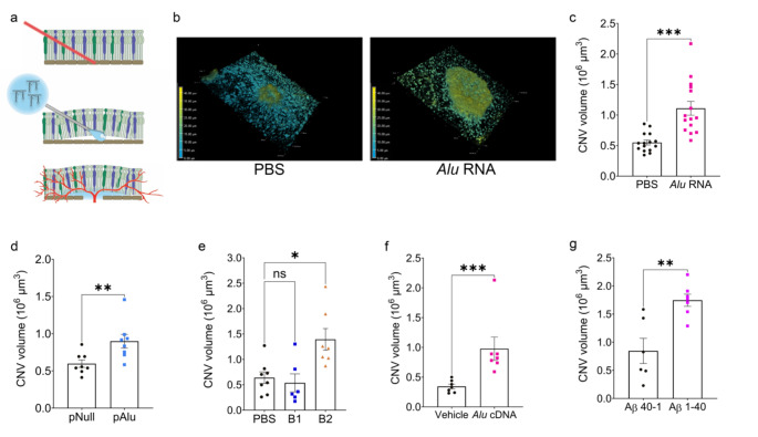
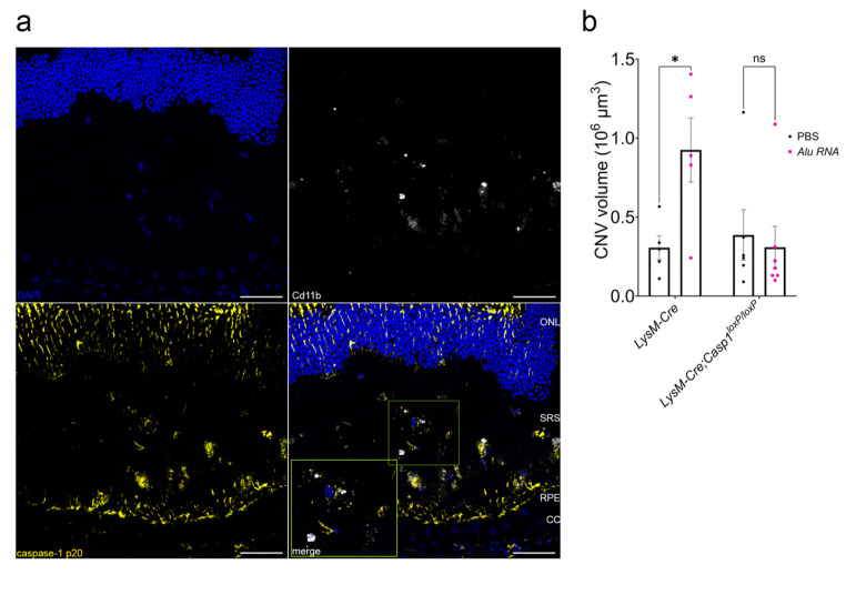
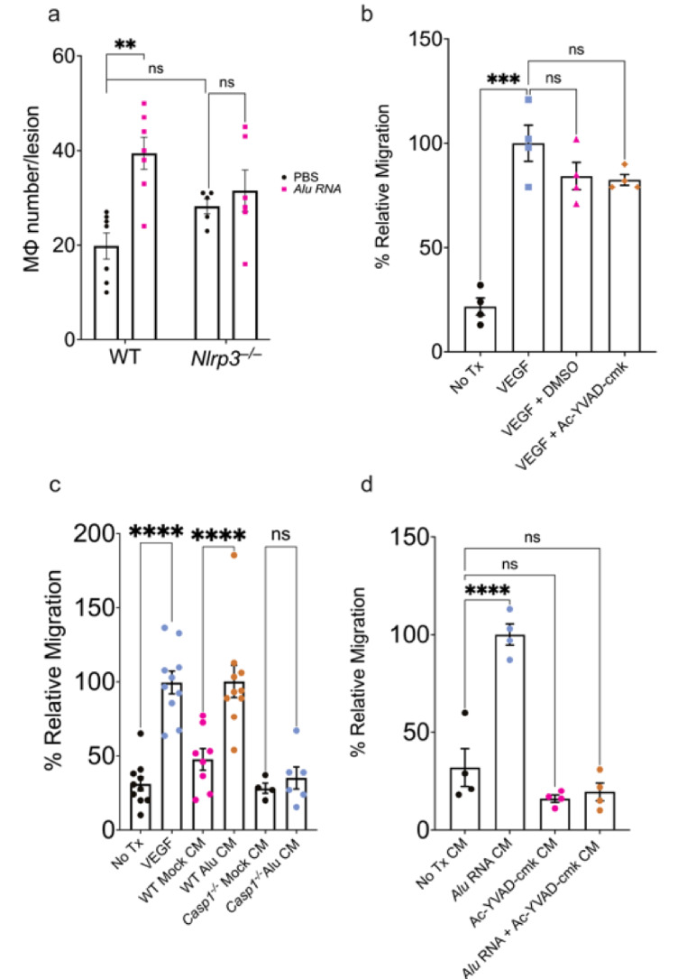
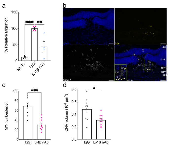
Inflammasome activation is implicated in diseases of aberrant angiogenesis such as age-related macular degeneration (AMD), though its precise role in choroidal neovascularization (CNV), a characteristic pathology of advanced AMD, is ill-defined. Reports on inhibition of inflammasome constituents on CNV are variable and the precise role of inflammasome in mediating pathological angiogenesis is unclear. Historically, subretinal injection of inflammasome agonists alone has been used to investigate retinal pigmented epithelium (RPE) degeneration, while the laser photocoagulation model has been used to study pathological angiogenesis in a model of CNV. Here, we report that the simultaneous introduction of any of several disease-relevant inflammasome agonists (Alu or B2 RNA, Alu cDNA, or oligomerized amyloid β (1-40)) exacerbates laser-induced CNV. These activities were diminished or abrogated by genetic or pharmacological targeting of inflammasome signaling constituents including P2rx7, Nlrp3, caspase-1, caspase-11, and Myd88, as well as in myeloid-specific caspase-1 knockout mice. Alu RNA treatment induced inflammasome activation in macrophages within the CNV lesion, and increased accumulation of macrophages in an inflammasome-dependent manner. Finally, IL-1β neutralization prevented inflammasome agonist-induced chemotaxis, macrophage trafficking, and angiogenesis. Collectively, these observations support a model wherein inflammasome stimulation promotes and exacerbates CNV and may be a therapeutic target for diseases of angiogenesis such as neovascular AMD.

Keywords: Age-related macular degeneration; Choroidal neovascularization; Inflammasome; Interleukin-1beta; Macrophage; Myd88.


Conflict of interest statement

Declarations Competing interests J.A: co-founder of DiceRx, iVeena Holdings, iVeena Delivery Systems and Inflammasome Therapeutics, and, unrelated to this work, has been a consultant for Abbvie/Allergan, Boehringer-Ingelheim, Janssen, Olix Pharmaceuticals, Retinal Solutions, and Saksin LifeSciences. B.D.G: co-founder of DiceRx. J.A., S.W., B.D.G.: inventors on matter-related patent applications filed by the University of Virginia or Kentucky.

Figures

Fig. 1
Fig. 1
Inflammasome agonism immediately following laser injury increases CNV volume. (a) Schematic demonstrating the combined laser CNV and subretinal injection (SRI) model. First, a laser burn is applied to rupture Bruch’s membrane. Subretinal injection is performed immediately following laser injury at the same site, and neovascularization begins to form around day 3. (b) Representative depth-coded 3D projections of laser CNV with SRI of PBS (left) or Alu RNA (right). Dimensions: 633.25 μm x 633.25 μm x 46 μm (c) CNV volumes quantified 7 days after combined laser injury and SRI of Alu RNA (P = 0.0001, Mann-Whitney test. N = 15 per group). (d) CNV volumes quantified 7 days after combined laser injury and in vivo transfection of plasmid-encoded Alu via SRI (P < 0.01, Mann-Whitney test. N = 8 per group). (e) CNV volumes quantified 7 days after combined laser injury and SRI of PBS, B1 (P > 0.99 vs. PBS), or B2 RNA (P = 0.03 vs. PBS, Kruskal-Wallis test. N = 6 (B1), N = 7 (B2)). (f) CNV volumes quantified 7 days after combined laser injury and SRI of Alu cDNA (P = 0.01, Mann-Whitney test. N = 7 (vehicle), N = 7 (Alu cDNA)). (g) CNV volumes quantified 7 days after combined laser injury and SRI of Aβ (P < 0.01, Mann-Whitney test. N = 6 (Aβ40−1), N = 7 (Aβ1−40))
Fig. 2
Fig. 2
Intact NLRP3 inflammasome components are required for inflammasome agonism-dependent CNV exacerbation. (a) CNV volumes quantified 7 days after combined laser injury and SRI of Alu RNA in P2rx7–/– mice (P > 0.99, Mann-Whitney test. N = 7–8). (b) CNV volumes quantified 7 days after combined laser injury, SRI of Alu RNA, and intravitreous pretreatment with PBS (P < 0.01), AZT (P = 0.20), or K8 (P = 0.50) (two-way ANOVA. N = 6 per group). (c) Quantification of CNV volume 7 days post Alu RNA SRI in Nlrp3–/– mice (P = 0.412, Mann-Whitney test. N = 11 (PBS), N = 9 (Alu RNA)). (d) CNV volumes quantification 7 days after laser injury and SRI of Alu RNA in Aim2–/– mice (P < 0.01, Mann-Whitney test. N = 6 per group). (e) CNV volumes quantified after combined laser injury and SRI of Alu RNA in Casp1/11–/– (P = 0.25), Casp11–/– (P = 0.25), and Casp1/11–/– x Casp11Tg+ (P > 0.99) (two-way ANOVA, N ≥ 5 per group). (f) CNV volume quantification 7 days after combined laser injury, intravitreous administration of either control peptide Z-FA-FMK (P < 0.01) or caspase-1 inhibitor Z-WEHD-FMK (P = 0.98), and Alu RNA SRI (two-way ANOVA, N = 6 per group). (g) CNV volumes quantified after 7 days post-laser injury and Alu RNA SRI in Myd88–/– mice (P = 0.79, Mann-Whitney U test, N ≥ 5 per group). (h) CNV volumes quantified 7 days after combined laser injury, intravitreous administration of a peptide MyD88 inhibitor (P = 0.08) or control peptide (P < 0.01), and Alu RNA SRI (P = 0.08, two-way ANOVA, N ≥ 6 per group)
Fig. 3
Fig. 3
Inflammasome activation in myelomonocytic cells is crucial to laser CNV. (a) Representative immunofluorescence images of cross-section of mouse retinae treated with Alu RNA. Slides were stained with indicated antibodies. Scale bars: 50 μm. CC: choriocapillaris; RPE: retinal pigmented epithelium; SRS: subretinal space; ONL: outer nuclear layer (b) CNV volume quantification 7 days post laser injury and Alu RNA SRI in LysM-Cre (P = 0.03, N = 5) and Casp1f/f x LysM-Cre (P = 0.91, N = 6) mice (two-way ANOVA)
Fig. 4
Fig. 4
Inflammasome activation promotes chemotaxis in peripheral BMDM. (a) Macrophage number quantification after 3 days post laser injury and Alu RNA SRI in WT (P = 0.03, N = 7) and Nlrp3–/– (P = 0.85, N = 7, two-way ANOVA). (b) Relative migration of WT BMDM toward the following chemoattractants: VEGF, VEGF + DMSO, VEGF + Ac-YVAD-cmk (P < 0.001 compared to untreated cells, N = 4, ordinary one-way ANOVA with Tukey’s multiple comparisons test). (c) Relative migration quantification of Alu RNA-transfected WT BMDM conditioned media (P < 0.01, N ≥ 8) and Alu RNA-transfected Casp1–/– BMDM (P = 0.97, N ≥ 4, ordinary one-way ANOVA with Tukey’s multiple comparisons test). (d) Relative migration of WT BMDM with the following conditioned media as chemoattractant: Alu RNA-transfected WT BMDM (P < 0.01); untransfected WT BMDM pretreated with Ac-YVAD-cmk (P = 0.24); Ac-YVAD-cmk pretreated, Alu RNA-transfected WT BMDM (P = 0.43, N = 4, ordinary one-way ANOVA with Tukey’s multiple comparisons test)
Fig. 5
Fig. 5
IL-1β neutralization reduces Alu RNA-induced chemotaxis, macrophage accumulation, and laser CNV exacerbation. (a) Relative migration of Alu RNA-transfected WT BMDM conditioned media pretreated with either IgG (P < 0.01) or IL-1β neutralizing antibody (P < 0.01, N = 4, ordinary one-way ANOVA with Tukey’s multiple comparisons test). (b) Representative images of Alu RNA-treated laser CNV lesions hybridized with probes against Il1b and Adgre1. Scale bar: 50 μm. CC: choriocapillaris; RPE: retinal pigmented epithelium; SRS: subretinal space; ONL: outer nuclear layer; INL: inner nuclear layer (c) Macrophage number and (d) CNV volume quantification after 3 days post laser injury and Alu RNA subretinal injection with either 500 ng IgG1 or IL-1β neutralizing antibody (P < 0.001, N = 8–10)

References

    1. Dridi S, Hirano Y, Tarallo V, Kim Y, Fowler BJ, Ambati BK et al (2012) ERK1/2 activation is a therapeutic target in age-related macular degeneration. Proc Natl Acad Sci 109(34):13781–13786 - PMC - PubMed
    1. Tarallo V, Hirano Y, Gelfand BD, Dridi S, Kerur N, Kim Y et al (2012) DICER1 loss and Alu RNA induce age-related macular degeneration via the NLRP3 inflammasome and MyD88. Cell 149(4):847–859 - PMC - PubMed
    1. Gelfand BD, Wright CB, Kim Y, Yasuma T, Yasuma R, Li S et al (2015) Iron Toxicity in the retina requires Alu RNA and the NLRP3 inflammasome. Cell Rep 11(11):1686–1693 - PMC - PubMed
    1. Kim Y, Tarallo V, Kerur N, Yasuma T, Gelfand BD, Bastos-Carvalho A et al (2014) DICER1/Alu RNA dysmetabolism induces caspase-8-mediated cell death in age-related macular degeneration. Proc Natl Acad Sci U S A 111(45):16082–16087 - PMC - PubMed
    1. Marneros AG (2013) NLRP3 inflammasome blockade inhibits VEGF-A-induced age-related macular degeneration. Cell Rep 4(5):945–958 - PMC - PubMed

LinkOut - more resources