. 2024 Nov 1;134(21):e175435.
doi: 10.1172/JCI175435.

Modeling primary microcephaly with human brain organoids reveals fundamental roles of CIT kinase activity

Affiliations

Modeling primary microcephaly with human brain organoids reveals fundamental roles of CIT kinase activity

Gianmarco Pallavicini et al. J Clin Invest. .

Abstract

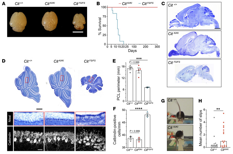
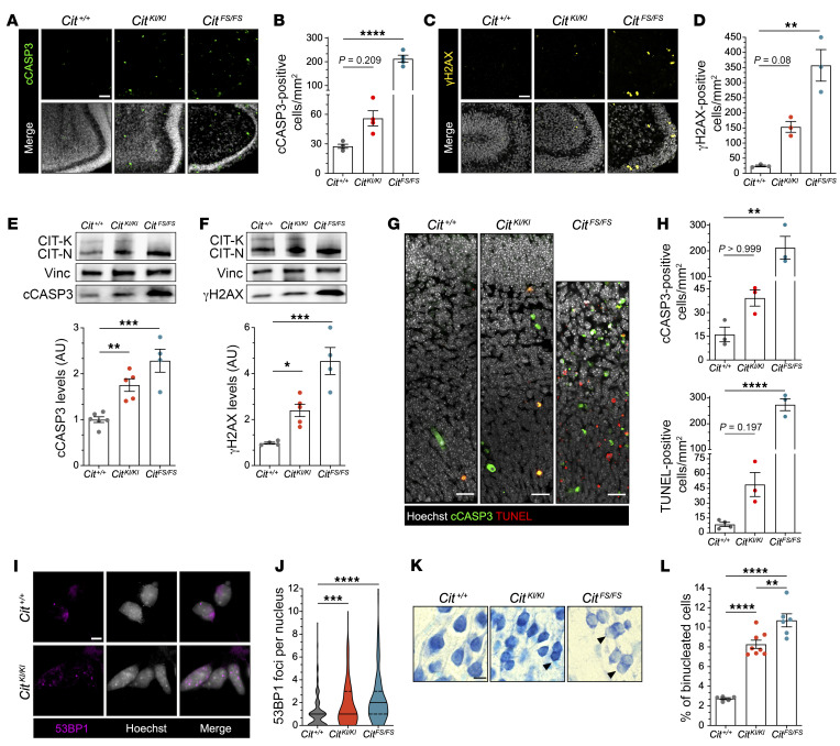
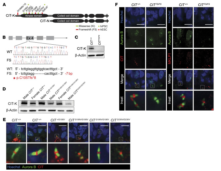
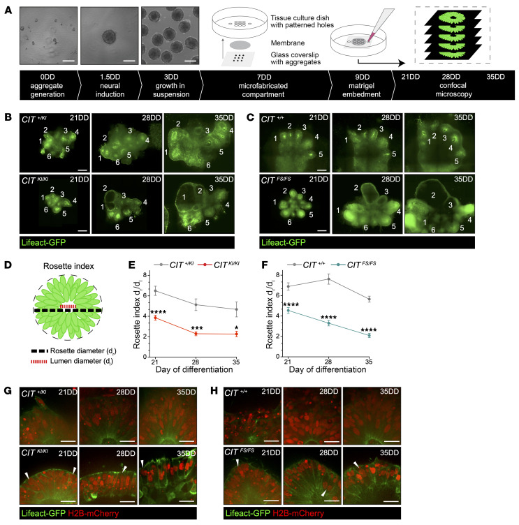
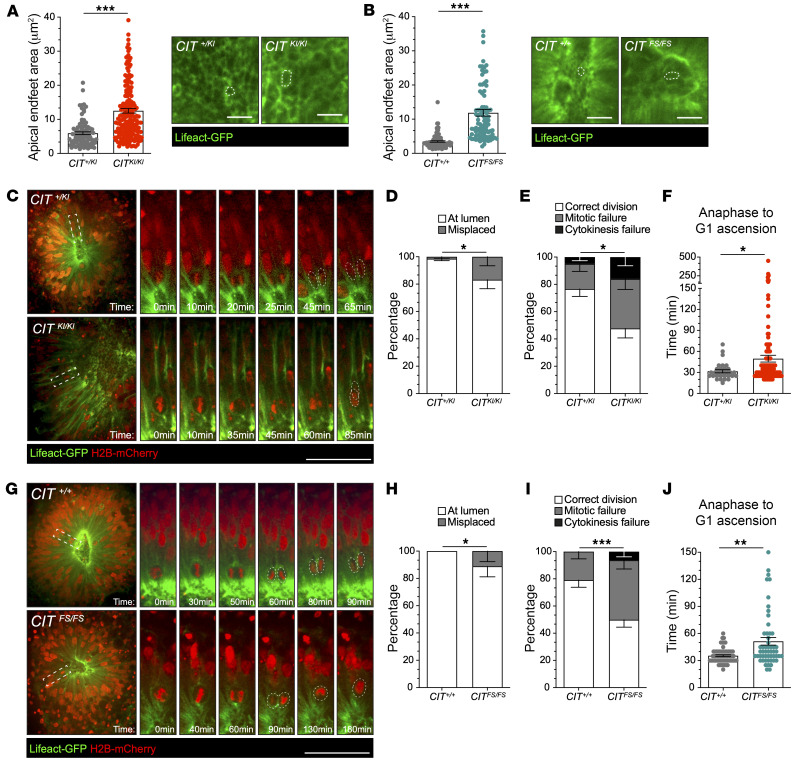
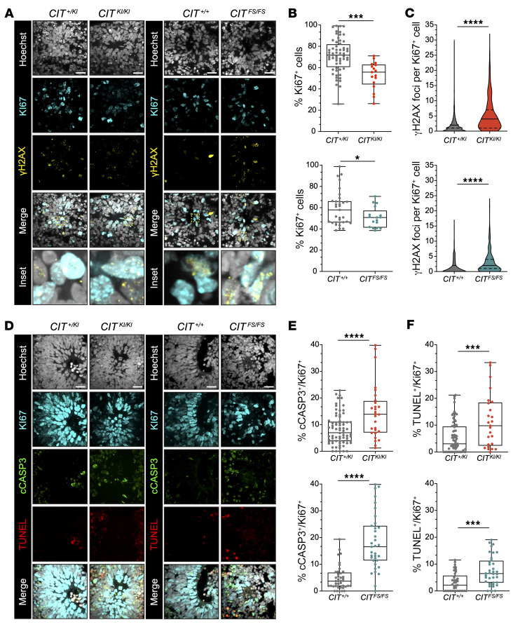
Brain size and cellular heterogeneity are tightly regulated by species-specific proliferation and differentiation of multipotent neural progenitor cells (NPCs). Errors in this process are among the mechanisms of primary hereditary microcephaly (MCPH), a group of disorders characterized by reduced brain size and intellectual disability. Biallelic citron rho-interacting serine/threonine kinase (CIT) missense variants that disrupt kinase function (CITKI/KI) and frameshift loss-of-function variants (CITFS/FS) are the genetic basis for MCPH17; however, the function of CIT catalytic activity in brain development and NPC cytokinesis is unknown. Therefore, we created the CitKI/KI mouse model and found that it did not phenocopy human microcephaly, unlike biallelic CitFS/FS animals. Nevertheless, both Cit models exhibited binucleation, DNA damage, and apoptosis. To investigate human-specific mechanisms of CIT microcephaly, we generated CITKI/KI and CITFS/FS human forebrain organoids. We found that CITKI/KI and CITFS/FS organoids lost cytoarchitectural complexity, transitioning from pseudostratified to simple neuroepithelium. This change was associated with defects that disrupted the polarity of NPC cytokinesis, in addition to elevating apoptosis. Together, our results indicate that both CIT catalytic and scaffolding functions in NPC cytokinesis are critical for human corticogenesis. Species differences in corticogenesis and the dynamic 3D features of NPC mitosis underscore the utility of human forebrain organoid models for understanding human microcephaly.

Keywords: Cell biology; Genetic diseases; Neurodevelopment; Neuronal stem cells; Neuroscience.


Figures

Figure 1
Figure 1. CitKI/KI mice show grossly normal CNS morphological structure with an ataxic phenotype.
(A) Representative image of Cit+/+, CitKI/KI, and CitFS/FS P10 brains. Scale bar: 5 mm. (B) Kaplan-Meier survival curves for CitKI/KI mice versus CitFS/FS (n = 12) mice. The log-rank (Mantel-Cox) test was used to compare survival between experimental groups (****P < 0.0001). (C) Cresyl Violet staining of sagittal sections of Cit+/+, CitKI/KI, and CitFS/FS P10 brains. Scale bar: 1 mm. (D) Upper panel: Cresyl violet staining of sagittal sections from the midline (vermis) of Cit+/+, CitKI/KI, and CitFS/FS P10 cerebella showing the anterior, central, and posterior sectors; scale bar: 500 μm. Middle panel: High-magnification Nissl stain of lobule V inset from the midline (vermis); scale bar: 50 μm. Lower panel: Immunofluorescence analysis for the Purkinje Cell marker calbindin 1 of sagittal sections of lobule V from the midline (vermis) obtained from P10 mice; scale bar: 25 μm. (E) Quantification of Purkinje cell layer (PCL) perimeter in the vermis of P10 Cit+/+, CitKI/KI, and CitFS/FS mice. (F) Quantification of Purkinje cell density per millimeter in the vermis of Cit+/+, CitKI/KI, and CitFS/FS P10 mice. Purkinje cells were stained for calbindin 1. (G) Representative picture extracted from videos of Cit+/+ and CitKI/KI mice slipping during beam walking test. (H) Quantification of the mean number of slips for 3 consecutive days of the beam-walking test on the same animal. Data indicate the mean ± SEM. **P < 0.01, ***P < 0.001, and ****P < 0.0001, by 1-way ANOVA followed by Bonferroni post hoc analysis (E and F) and unpaired, 2-tailed Student’s t test (H). Each dot represents an independent animal.
Figure 2
Figure 2. CitKI/KI mice show increased apoptosis, DNA damage, and cytokinesis failure in CNS tissue, but less than CitFS/FS mice.
(A) Immunofluorescence analysis of P4 cerebella from mice of the indicated genotypes for the apoptotic marker cCASP3 (green) and Hoechst (gray). Scale bar: 25 μm. (B) Quantification of cCASP3+ cells in A. (C) Immunofluorescence analysis of P4 cerebella from mice of the indicated genotypes for the DNA damage marker γH2AX (yellow) and Hoechst (gray). Scale bar: 25 μm. (D) Quantification of γH2AX+ cells in C. (E and F) Western blot analysis of total lysate from P4 cerebella from mice of the indicated genotypes. The levels of cCASP3 (E) and γH2AX (F) were analyzed relative to the internal loading control vinculin (Vinc). Each dot represents an independent replicate. (G) Immunofluorescence analysis for the apoptotic marker cCASP3 (green), TUNEL assay (red), and Hoechst (gray) of E14.5 cortex obtained from embryos of the indicated genotypes. Scale bars: 10 μm. (H) Quantification of cCASP3 and TUNEL+ cells in (G). (I) Representative images of E12.5 NPCs from Cit+/+ and Cit KI/KI embryos, immunostained for 53BP1 (magenta) and Hoechst (gray) 18 hours after plating. Scale bar: 10 μm. (J) Quantification of 53BP1 nuclear foci in I; more than 250 cells were counted for each genotype in each replicate (n = 3). (K) High-power fields of Cresyl violet–stained coronal sections of P10 cortex obtained from mice of the indicated genotypes. Arrowheads indicate binucleated cells. Scale bar: 10 μm. (L) Quantification of binucleated cells in K. In microscopy quantifications, every dot represents an independent animal and at least 9 fields per genotype were analyzed. Data indicate the mean ± SEM. *P < 0.05, **P < 0.01,***P < 0.001, and ****P < 0.0001, by 1-way ANOVA followed by Holm-Šidák post hoc analysis.
Figure 3
Figure 3. Modeling CIT variants using human models of neurodevelopment.
(A) Scheme of CIT-K and CIT-N protein isoforms. Homozygous and compound heterozygous variants localized in the N-terminus and within the kinase domain of CIT-K. Missense variants are depicted in green, and LOF FS and splice (SpEx) variants are depicted in red. (B) CRISPR/Cas9-mediated targeting of the CIT locus at exon 4. Sequences and chromatograms of the 7 bp deletion are shown, with the corresponding alteration of the protein sequence highlighted in red. (C) Western blot analysis of CIT expression in NPCs of the indicated genotypes. The CIT-K isoform was absent in the CRISPR/Cas9-edited CITFS/FS line. (D) Western blot analysis of CIT expression in NPCs of the indicated genotypes. Variability in CIT-K abundance was detected in CITKI/KI NPCs compared with unaffected controls. (E) Immunofluorescence of Aurora B (green) midbody arms and CIT (red) show the presence of CIT in the midbody central bulge in the indicated genotypes of 35DD dorsal forebrain organoids. DNA stained with Hoechst (blue). Scale bars: 10 μm. (F) Immunofluorescence of Aurora B (green) midbody arms and the midbody central bulge markers CIT (red) and MKLP1 (red) in 35DD dorsal forebrain organoids. DNA stained with Hoechst (blue). CIT was absent in the midbody central bulge in CITFS/FS 35DD organoids. Immunofluorescence with the midbody central bulge marker MKLP1 (red) shows the presence of this structure flanked by Aurora B (green) midbody arms in CITFS/FS 35DD organoids. Scale bars: 10 μm.
Figure 4
Figure 4. CIT-affected organoids demonstrate changes to pseudostratified neuroepithelium.
(A) Schematic of dorsal forebrain organoid differentiation and microfabricated compartment generation with the corresponding time stamps. Neural differentiation begins with hPSC aggregate generation at 0DD, followed by neural induction at 1.5DD. An illustration of the microfabricated compartment depicts the orientation of compartment components. Neural aggregates are positioned on glass coverslips in a 3 × 3 pattern at 7DD, and the compartment is sealed with a UV adhesive. Neural differentiation medium fills the tissue culture dish to facilitate medium exchange to the compartment containing the developing tissue. Two days later, on 9DD, the compartment is embedded with Matrigel. Inverted confocal microscopy is performed on 21DD, 28DD, and 35DD. Scale bars: 200 μm. (B and C) Representative images of developing organoids and rosettes (white numbers) across time using Lifeact-GFP. Affected CITFS/FS and CITKI/KI rosettes exhibited large lumens and a reduction in neuroepithelial complexity. Scale bars: 250 μm. (D) Illustration of rosette measurements performed across CIT organoids, with diameter measurements and Ri calculations across organoid rosettes. (E and F) Quantification of Ri (dr/dl) in CIT+/KI and in CITKI/KI organoids (E) and CIT+/+ and in CITFS/FS organoids (F). (G and H) Representative insets of developing rosettes across time using Lifeact-GFP and H2B-mCherry. Control rosettes maintained a pseudostratified neuroepithelium at all 3 time points, while many affected CITKI/KI and CITFS/FS rosettes showed transition toward a simple epithelial architecture. Multinucleated cells were apparent in the rosette, and examples are shown (white arrowheads). Scale bars: 25 μm. Quantification was done from a minimum of 2 independent compartment preparations per genotype. Each compartment contained 9 or fewer organoids per preparation. Data indicate the mean ± SEM. *P < 0.05, ***P < 0.001, and ****P < 0.0001, by repeated-measures, 2-way ANOVA.
Figure 5
Figure 5. CIT-affected organoids show aNPC apical endfeet surface area increases and mitotic defects.
(A and B) Apical endfeet surface area and representative images of 35DD organoid apical surface labeled with Lifeact-GFP in microfabricated compartments. The apical endfeet surface area was increased in affected CITKI/KI (A) and CITFS/FS (B) organoids compared with CIT+/KI and CIT+/+ control organoids. Scale bars: 10 μm. (C) Representative images from videos of CIT+/KI and CITKI/KI rosettes with aNPC mitotic division (white dashed rectangles) at the apical surface. Panel shows the time details of this division. Scale bar: 50 μm. (D) Quantification of the percentage of division occurring at the central lumen or misplaced from the lumen in C. (E) Quantification of the percentage of correct division, mitotic failure, and cytokinesis failure in C. (F) Measurement of the time spent from anaphase to G1 ascension in CIT+/KI and CITKI/KI aNPC divisions in 35DD organoid rosettes. Each dot indicates a single dividing cell. (G) Representative images from videos of CIT+/+ and CITFS/FS rosettes with aNPC mitotic division (white dashed rectangles) at the apical surface. Panel shows the time details of this division. Scale bar: 50 μm. (H) Quantification of the percentage of division occurring at the central lumen or misplaced from the lumen in G. (I) Quantification of the percentage of correct division, mitotic failure, and cytokinesis failure in G. (J) Measurement of the time spent from anaphase to G1 ascension in CIT+/+ and CITFS/FS aNPC divisions in 35DD organoid rosettes. Each dot indicates a single dividing cell. Quantification was done from a minimum of 2 independent compartment preparations per genotype. The compartments each contained 9 or fewer organoids per preparation. Data indicate the mean ± SEM. *P < 0.05, **P < 0.01, and ***P < 0.001, by unpaired, 2-tailed Student’s t test for the apical endfeet area (A and B), Fisher’s exact probability test for the percentage distribution (D, E, H, and I), and Mann-Whitney U test for the time from anaphase to G1 ascension (F and J).
Figure 6
Figure 6. CIT-affected organoids show accumulation of DNA damage and apoptosis.
(A) Representative images of rosettes for the indicated CIT genotypes for the DNA damage marker γH2AX (yellow), the proliferation marker Ki67 (cyan), and Hoechst (gray). Scale bars: 20 μm. (B and C) Quantification of Ki67+ cells (B) and γH2AX foci per Ki67+ cell (C). More than 500 cells were counted for each genotype in each replicate. (D) Representative images of rosettes for the indicated CIT genotypes for the apoptotic marker cCASP3 (green), TUNEL (red), the proliferation marker Ki67 (cyan), and Hoechst (gray). Scale bars: 20 μm. (E and F) Quantification of cCASP3 (E) and TUNEL (F) staining per Ki67+ cells. Every dot represents a neural rosette. Quantification was done from a minimum of 3 independent organoid preparations. Data indicate the mean ± SEM. *P < 0.05, ***P < 0.001, and ****P < 0.0001, by unpaired, 2-tailed Student’s t test (B, E, and F) and Mann-Whitney U test (for γH2AX foci in C).

References

    1. Vaid S, Huttner WB. Progenitor-based cell biological aspects of neocortex development and evolution. Front Cell Dev Biol. 2022;10:892922. doi: 10.3389/fcell.2022.892922. - DOI - PMC - PubMed
    1. Miller DJ, et al. Shared and derived features of cellular diversity in the human cerebral cortex. Curr Opin Neurobiol. 2019;56:117–124. doi: 10.1016/j.conb.2018.12.005. - DOI - PMC - PubMed
    1. Villalba A, et al. The regulation of cortical neurogenesis. Curr Top Dev Biol. 2021;142:1–66. doi: 10.1016/bs.ctdb.2020.10.003. - DOI - PubMed
    1. Espinós A, et al. Evolution of genetic mechanisms regulating cortical neurogenesis. Dev Neurobiol. 2022;82(5):428–453. doi: 10.1002/dneu.22891. - DOI - PMC - PubMed
    1. Götz M, Huttner WB. The cell biology of neurogenesis. Nat Rev Mol Cell Biol. 2005;6(10):777–788. doi: 10.1038/nrm1739. - DOI - PubMed

MeSH terms

Substances