Review
. 2025 Apr;31(2):195-214.
doi: 10.1177/10738584241275583. Epub 2024 Sep 24.

The effect of traumatic brain injury on learning and memory: A synaptic focus

Affiliations
Review

The effect of traumatic brain injury on learning and memory: A synaptic focus

Eric Eyolfson et al. Neuroscientist. 2025 Apr.

Abstract

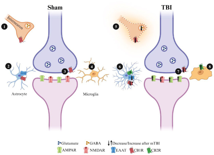
Deficits in learning and memory are some of the most commonly reported symptoms following a traumatic brain injury (TBI). We will examine whether the neural basis of these deficits stems from alterations to bidirectional synaptic plasticity within the hippocampus. Although the CA1 subregion of the hippocampus has been a focus of TBI research, the dentate gyrus should also be given attention as it exhibits a unique ability for adult neurogenesis, a process highly susceptible to TBI-induced damage. This review examines our current understanding of how TBI results in deficits in synaptic plasticity, as well as how TBI-induced changes in endocannabinoid (eCB) systems may drive these changes. Through the synthesis and amalgamation of existing data, we propose a possible mechanism for eCB-mediated recovery in synaptic plasticity deficits. This hypothesis is based on the plausible roles of CB1 receptors in regulating inhibitory tone, influencing astrocytes and microglia, and modulating glutamate release. Dysregulation of the eCBs may be responsible for deficits in synaptic plasticity and learning following TBI. Taken together, the existing evidence indicates eCBs may contribute to TBI manifestation, pathogenesis, and recovery, but it also suggests there may be a therapeutic role for the eCB system in TBI.

Keywords: dentate gyrus; electrophysiology; endocannabinoids; hippocampus; long-term depression; long-term potentiation; synaptic plasticity.


Conflict of interest statement

Declaration of Conflicting InterestsThe author(s) declared no potential conflicts of interest with respect to the research, authorship, and/or publication of this article.

Figures

Box 1.
Box 1.
Introduction to field electrophysiology. (A) Major hippocampal pathways that compose the trisynaptic loop. The dentate gyrus receives both feed-forward and feed-back inhibition from local interneurons. In vitro electrophysiology necessitates a GABA-A antagonist to block feed-forward inhibition. Primary recording pathways for in vitro field electrophysiology include the medial and lateral performant pathways in the dentate gyrus and Schaffer collateral pathways in CA1. (B) Brightfield micrograph (4× magnification) of transverse hippocampal tissue recording in the medial perforant pathway of the dentate gyrus. It illustrates the placement of a concentric bipolar stimulating electrode (right) and glass recording pipette (left). (C) Sample waveform characteristic of in vitro field electrophysiology experiments: (i) Stimulation artifact, indicating the conduction of electricity through the artificial cerebral spinal fluid (aCSF) medium. (ii) Fiber volley, signifying presynaptic neurotransmitter release as electricity moves from the stimulating to the recording electrode. (iii) Presynaptic neurotransmitter release activates postsynaptic receptors, initiating a postsynaptic excitatory response, initially depolarized by AMPA receptors and subsequently NMDA receptors. To gauge synaptic communication strength, the initial slope of the field excitatory post-synaptic potential (fEPSP) is measured. (D) Graphical representation of the slope of the fEPSP denoted as response size across time. Stable baseline recordings (typically a minimum of 20 minutes) precede evoke responses. Following the baseline period, high-frequency stimulation (100 Hz/1 s) can induce an increase the size of the responses (known as long-term potentiation), while low-frequency stimulation (1 Hz/15 min) can decrease response size (known as long-term depression). (E) Sample traces of hippocampal slices subjected to high-frequency or low-frequency stimulation, presented as a percentage of the baseline recording. Each form of bidirectional synaptic plasticity exhibits a significant initial increase (+125% in this HFS induction) or decrease (–100% in this low-frequency stimulation induction) in response size that eventually stabilizes. The recording duration is generally 60 minutes following high- or low-frequency stimulation as the mechanisms for LTP and LTD transition into protein-dependent phases.
Figure 1.
Figure 1.
Simplified diagram of steps involved in the induction of a postsynaptically mediated form of long-term potentiation induction at hippocampal excitatory synapses. Excitatory glutamatergic synapses normally possess a complement of AMPA and NMDA receptors in the postsynaptic terminal. The administration of (1) high-frequency (i.e., 100 Hz) conditioning stimulation causes the release of glutamate (2), which primarily activates AMPARs initially (3). In the dentate gyrus (DG) (4), GABAergic synapses can also play a significant role in the induction of LTP in the medial perforant path synapses, and LTP induction normally requires blocking feedforward inhibition with GABA-A antagonists (i.e., bicuculine or picrotoxin). When inhibition is blocked, the activation of AMPARs results in an influx of Na+ into the postsynaptic neuron, depolarizing the synapse and alleviating the MG2+ block of the NMDAR pore. (5) The NMDARs can now influx Ca2+ into the postsynaptic cell, and this activates calmodulin (CaM) and calcium-calmodulin dependent protein kinase II (CamKII). CaM also activates adenyl cyclase, which results in the production of cyclic adenosine monophosphate (cAMP) and then activation of protein kinase A (PKA) by cAMP. Kinases, such as CamKII and PKA, phosphorylate GluRs, leading to the insertion of additional AMPARs into the postsynaptic membrane. CamKII and cAMP also activate mitogen-activated protein kinase A, which is translocated to the nucleus and activates cAMP response binding element-binding protein (CREB). CREB is a transcription factor and binds to the CRE response element, leading to the transcription of genes necessary for long-lasting protein synthesis-dependent forms of LTP.
Figure 2.
Figure 2.
Simplified diagram of long-term depression induction in hippocampal excitatory synapses. (1) Induction of a low-frequency stimulation leads to modest depolarization, which (2) releases glutamate and activates AMPARs. (3) Activated AMPARs results in an influx of Na+ into the postsynaptic neuron but not enough to fully clear the MG2+ block of the NMDAR pore. (4) Influx of Ca2+ results in the activation of CaM, which leads to the activation of calcineurin. Calcineurin activates phosphatases such as protein phosphatase 1, leading to the dephosphorylation and endocytosis of AMPARs.
Figure 3.
Figure 3.
Deficits in long-term potentiation are seen in adolescent female animals following three repetitive injuries (72-hour interinjury interval) in the medial perforant pathway of the dentate gyrus. (A) Time course of synaptic plasticity experiments presented as a percentage of the baseline fEPSP. The black arrow indicates high-frequency stimulation (HFS) induction (4 × 100 Hz). Short-term plasticity is measured as the change in response size 1 minute following HFS. Long-term potentiation is measured as the change in response size 55 to 60 minutes following HFS. (B) Long-term potentiation is decreased following repetitive mild traumatic brain injury with sample traces (C) from sham and repetitive mild traumatic brain injury hippocampal slices. *P < 0.05 (Student’s t-test). Unpublished data. For more information on injury protocol, see Eyolfson and others (2021). For more information on electrophysiologic methods, see Fontaine and others (2019).
Figure 4.
Figure 4.
CB1R immunolocalization in the molecular layer of the dentate gyrus. Electron microscopy of CB1R immunogold particles (black and white arrows) is localized to inhibitory (ter, light green) and excitatory synaptic terminals (ter, blue) as well as to astrocytes (as, dark green) and mitochondria (m, purple). Postsynaptic dendrites (den, pink) and dendritic spines (sp, pink) are CB1R immunonegative. For more information on electron microscopy methodology, see Bonilla-Del Río and others (2021).
Figure 5.
Figure 5.
Simplified graphic displaying the potential roles CB1 receptors could play in synaptic plasticity at excitatory hippocampal synapses. Cannabinoid receptors are located in a number of places where they have the potential to significantly affect synaptic plasticity. In response to injury, microglia are activated and proliferate as part of the proinflammatory response. Astrocytes also contribute to neuroinflammation by becoming reactive following injury. Both astrocytes and microglia synthesize endocannabinoids (eCBs) and express cannabinoid receptors (CBRs), which could potentially aid in decreasing neuroinflammation. (1/5) CB1 receptors located on presynaptic terminals that release GABA can reduce inhibitory tone when activated. This can lead to greater postsynaptic excitation and enhance the capacity for LTP at excitatory synapses. Following mild traumatic brain injury (mTBI), glutamate excitotoxicity results in decreased GABAergic functioning and interneuron cell death, further driving aberrant excitatory transmission. (2/6) CB1R located at astrocytes can reduce the uptake of glutamate, both increasing excitatory drive at a synapse and lowering the conversion of glutamate to GABA by astrocytes. Following injury, upregulation of CB2R on astrocytes can reduce proinflammatory responses and improve conditions for neuronal survival. (3/7) Retrograde transmission of eCBs synthesized from the postsynaptic terminal can interact with CB1Rs located on presynaptic terminals and modulate the release of glutamate. (4/8) CB2Rs located on microglia are downregulated under homeostatic conditions, and following mTBI, CB2R is upregulated to reduce proinflammatory responses.

Similar articles

Cited by

References

    1. Ahluwalia M, Mcmichael H, Kumar M, Espinosa MP, Bosomtwi A, Lu Y, and others. 2023. Altered endocannabinoid metabolism compromises the brain-CSF barrier and exacerbates chronic deficits after traumatic brain injury in mice. Exp Neurol 361:114320. - PMC - PubMed
    1. Albensi BC, Sullivan PG, Thompson MB, Scheff SW, Mattson MP. 2000. Cyclosporin ameliorates traumatic brain-injury-induced alterations of hippocampal synaptic plasticity. Exp Neurol 162(2):385–9. - PubMed
    1. Alkadhi KA. 2019. Cellular and molecular differences between area CA1 and the dentate gyrus of the hippocampus. Mol Neurobiol 56(9):6566–80. - PubMed
    1. Almeida-Suhett CP, Prager EM, Pidoplichko V, Figueiredo TH, Marini AM, and others. 2015. GABAergic interneuronal loss and reduced inhibitory synaptic transmission in the hippocampal CA1 region after mild traumatic brain injury. Exp Neurol 273:11–23. - PubMed
    1. Amenta PS, Jallo JI, Tuma RF, Elliott MB. 2012. A cannabinoid type 2 receptor agonist attenuates blood–brain barrier damage and neurodegeneration in a murine model of traumatic brain injury. J Neurosci Res 90(12):2293–305. - PubMed

MeSH terms

Substances