. 2024 Sep 24;19(9):e0308821.
doi: 10.1371/journal.pone.0308821. eCollection 2024.

Update on a brain-penetrant cardiac glycoside that can lower cellular prion protein levels in human and guinea pig paradigms

Affiliations

Update on a brain-penetrant cardiac glycoside that can lower cellular prion protein levels in human and guinea pig paradigms

Shehab Eid et al. PLoS One. .

Abstract

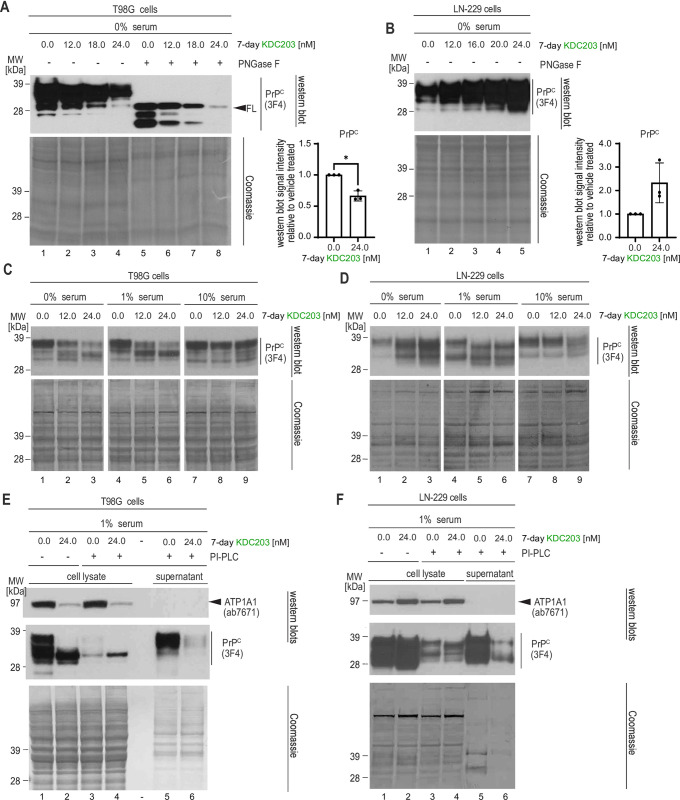
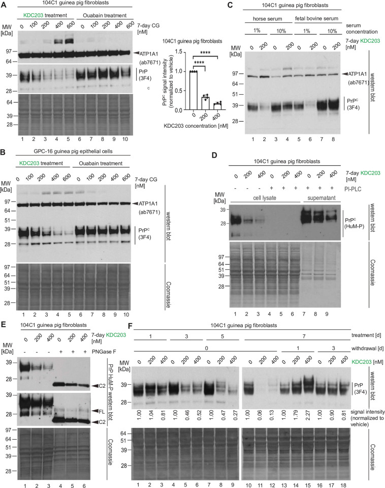
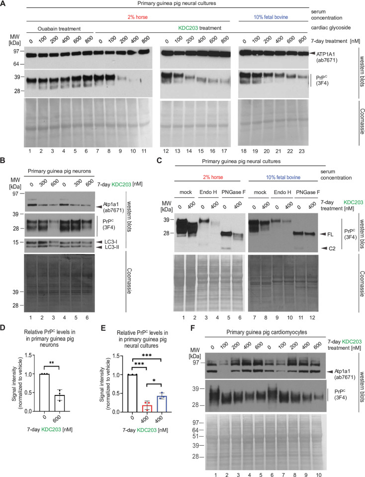
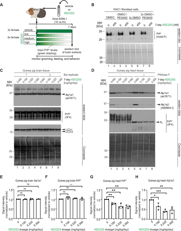
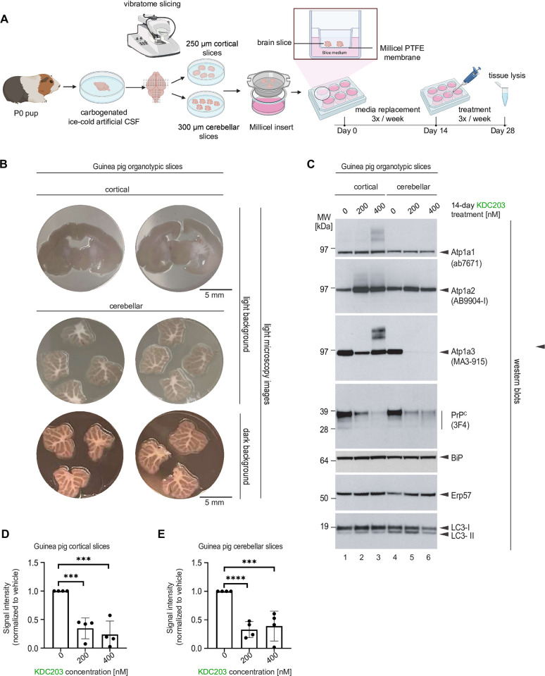
Lowering the levels of the cellular prion protein (PrPC) is widely considered a promising strategy for the treatment of prion diseases. Building on work that established immediate spatial proximity of PrPC and Na+, K+-ATPases (NKAs) in the brain, we recently showed that PrPC levels can be reduced by targeting NKAs with their natural cardiac glycoside (CG) inhibitors. We then introduced C4'-dehydro-oleandrin as a CG with improved pharmacological properties for this indication, showing that it reduced PrPC levels by 84% in immortalized human cells that had been differentiated to acquire neural or astrocytic characteristics. Here we report that our lead compound caused cell surface PrPC levels to drop also in other human cell models, even when the analyses of whole cell lysates suggested otherwise. Because mice are refractory to CGs, we explored guinea pigs as an alternative rodent model for the preclinical evaluation of C4'-dehydro-oleandrin. We found that guinea pig cell lines, primary cells, and brain slices were responsive to our lead compound, albeit it at 30-fold higher concentrations than human cells. Of potential significance for other PrPC lowering approaches, we observed that cells attempted to compensate for the loss of cell surface PrPC levels by increasing the expression of the prion gene, requiring daily administration of C4'-dehydro-oleandrin for a sustained PrPC lowering effect. Regrettably, when administered systemically in vivo, the levels of C4'-dehydro-oleandrin that reached the guinea pig brain remained insufficient for the PrPC lowering effect to manifest. A more suitable preclinical model is still needed to determine if C4'-dehydro-oleandrin can offer a cost-effective complementary strategy for pushing PrPC levels below a threshold required for long-term prion disease survival.


Conflict of interest statement

I have read the journal’s policy and the authors of this manuscript have the following competing interests: GS and DW declare that they are co-inventors on patent applications named ‘Compounds for altering levels of one or more NKA alpha subunits and their use in treating prion diseases or brain diseases associated with cellular prion protein’ filed in the US and Canada as well as a European patent application named ‘Compounds and methods to treat prion and related diseases’ based on a PCT international filing (PCT/CA2022/050358). These patent applications describe the use of CGs for the purpose of reducing steady-state levels of PrPC. This potential conflict-of-interest does not affect the adherence of all authors to journal policies on the sharing of data and materials. This does not alter our adherence to PLoS One policies on sharing data and materials.

Figures

Fig 1
Fig 1. The PrPC lowering potency of KDC203 affects all post-translational PrPC isoforms but is reduced in rich culture media.
(A) KDC203 concentration-dependent reduction of all PrPC isoforms is apparent in PNGase F treated human T98G cell extracts. The Coomassie stain documents equal loading. (B) Human LN-229 cells exhibit a KDC203 concentration-dependent increase in total PrPC levels. (C) The capacity of KDC203 to lower PrPC levels in T98G cells is reduced in serum-rich media. (D) The KDC203-dependent increase of PrPC levels in LN-229 levels is reduced in serum-rich media. (E) The cell surface pool of PrPC that can be released into the supernatant by PI-PLC digestion dominates total PrPC levels in T98G cells and is strongly reduced upon KDC203 treatment. ATP1A1 levels are also reduced in KDC203-treated T98G cells but cannot be observed in the PI-PLC digestion supernatant. (F) KDC203 treatment caused an increase in total ATP1A1 and PrPC levels in LN-229 cells yet reduced the PI-PLC-releasable cell surface pool of PrPC.
Fig 2
Fig 2. KDC203 treatment lowers PrPC levels in guinea pig cells and PrPC expression temporarily overshoots when KDC203 is withdrawn.
(A) The guinea pig cell line 104C1 which exhibits a fibroblast morphology responds to 7-day treatment with KDC203, but not Ouabain, with a significant reduction in its PrPC levels. However, the KDC203 concentrations required for this outcome exceed approximately 30-fold those observed to achieve a similar PrPC reduction in human cells. The asterisk indicates high molecular weight ATP1A1 antibody-reactive bands in a subset of fractions. Although the identity of these bands has not been resolved, their presence is consistent with partially SDS-stable ATP1A1-containing complexes. (B) The epithelial guinea pig cell line, GPC-16, resembles 104C1 cells in their response to KDC203 or Ouabain. A Coomassie stain of the western blot membrane validated equal protein loading. ATP1A1 signals were revealed following a reprobe of the PrPC blot. (C) Guinea pig 104C1 cells recapitulate reduced PrPC lowering potency of KDC203 when grown in media with higher serum levels. Note that the relatively stable ATP1A1 levels serve partially as a loading control as they were revealed on the same membrane following a reprobe of the PrPC blot. (D) Upon PI-PLC cleavage of guinea pig 104C1 cells, no PrPC can be detected in cellular lysates, and the PI-PLC releasable pool is diminished in KDC203 treated cells. (E) By its apparent molecular weight, PrPC expressed in guinea pig 104C1 cells is predominantly composed of the N-terminally truncated C2 isoform. (F) Daily KDC203 treatment of guinea pig 104C1 cells caused a time-dependent reduction of steady-state PrPC levels, culminating in a profound reduction after 7 days of treatment. The subsequent withdrawal of KDC203 reveals a temporary upregulation of the underlying PrPC expression whose magnitude correlates with the prior KDC203 concentration used to achieve the PrPC lowering effect. This effect ceases after 3 days of KDC203 withdrawal.
Fig 3
Fig 3. KDC203 lowers PrPC levels in primary cultures of guinea pig neurons, astrocytes, or cardiomyocytes.
(A) KDC203, but not Ouabain, lowers steady-state PrPC levels in primary guinea pig neural cultures, with the PrPC lowering potency enhanced in media containing 2% horse serum than 10% fetal bovine serum. (B) Primary neurons respond to KDC203 with the lowering of steady-state Atp1a1 and PrPC protein levels. LC3-directed western blotting does not reveal the molecular autophagy signature even when cells were exposed to 600 nM KCD203 levels. (C) 7-day KDC203 treatment of primary guinea pig astrocytes led to a reduction in steady-state PrPC levels and a downward shift in its apparent molecular weight that was more pronounced in 2% horse serum than 10% fetal bovine serum. The subsequent digestion with endoglycosidase H (Endo H) or PNGase F failed to reveal a shift in the apparent MW of PrPC in response to Endo H yet documented a profound shift upon PNGase F digestion, consistent with KDC203 at 400 nM having had no impact on the ability of PrPC to reach the cell surface. Note that this analysis did not consider relative signal intensities prior to and after digestion (likely caused by a reduced propensity of PrPC to transfer to the western blot transfer upon removal of its N-glycans). (D) Densitometric western blot quantitation of steady-state PrPC levels in primary guinea pig neurons following 7-day KDC203 treatment, including the results shown in Panel B. (E) Densitometric western blot quantitation of steady-state PrPC levels in primary guinea pig neural cell cultures that were for 7 days exposed to vehicle or 400 nM KDC203 in the presence of 2% horse serum (red bar) or 10% fetal bovine serum (blue bar), including the results shown in Panels A and C. (F) 7-day KDC203 treatment of primary guinea pig cardiomyocytes led to a bimodal shift in the steady-state levels of Atp1a1 that was most pronounced at 100 nM treatment concentrations. In contrast, the signal intensities of PrPC western blot bands declined in a KDC203 concentration-dependent manner that was accompanied by an increase in their mobility in samples derived from KDC203-treated cardiomyocytes. Two biological replicates were included for each KDC203 concentration to document the reproducibility of these complex changes to PrPC and Atp1a1 signals in this cell model.
Fig 4
Fig 4. Subcutaneous administration of KDC203 to guinea pigs lowered steady-state PrPC levels in the heart but not in brain tissue.
(A) Cartoon depicting design of guinea pig KDC203 treatment study. (B) Analysis of the impact of KDC203 formulation on its ability to lower steady-state PrPC levels in 104C1 guinea pig cells. (C) 7-day subcutaneous administration of KDC203 to guinea pigs did not change steady-state guinea pig brain Atp1a1 or PrPC levels or cause a molecular LC3 signature that indicated autophagy in brain tissue. (D) 7-day subcutaneous administration of KDC203 to guinea pigs lowered steady-state Atp1a1, Atp1a2 and PrPC protein levels in heart tissue. PNGase F digestion of extract proteins revealed that total PrPC levels in this tissue were predominantly comprised of full-length PrPC. (E-H) Subcutaneous 7-day administration of KDC203 in guinea pigs revealed no significant effect of KDC203 on the brain but a significant dose-dependent effect on the heart. Densitometric quantitation of steady-state brain and heart Atp1a1 levels (E, G) and PrPC levels (F, H).
Fig 5
Fig 5. KDC203 lowers PrPC levels in cultured guinea pig cortical and cerebellar brain slices.
(A) Cartoon depicting the workflow for generating cortical and cerebellar guinea pig organotypic brain slices. (B) Microscopy images of cortical and cerebellar guinea pig organotypic brain slices. (C) Western blot analyses of proteins-of-interest following 14-day treatment without or with 200 nM or 400 nM KDC203 in the brain-in-a-dish culture medium. Note that KDC203 exhibited inconsistent effects on the three NKA α subunits, with steady-state Atp1a1 levels being the least affected, Atp1a2 levels elevated in both cortical and cerebellar slices exposed to KDC203, and Atp1a3 levels being differently affected in cortical and cerebellar slices by the presence of KDC203. In contrast, steady-state PrP levels were consistently lower in a KDC203 concentration-dependent manner in both cortical and cerebellar organotypic brain slices. Finally, markers of endoplasmic reticulum stress, BiP and Erp57, or autophagy, LC3, were not significantly altered in the two types of brain slices with or without KDC203 exposure. (D, E) Quantitation of steady-state PrPC levels in vehicle-treated or KDC203-treated cortical (D) or cerebellar (E) brain slices. Significance thresholds indicated in the graphs are based on densitometric analyses of western blot signal intensities from four biological replicates for each treatment group.

References

    1. Minikel EV, Vallabh SM, Lek M, Estrada K, Samocha KE, Sathirapongsasuti JF, et al.. Quantifying prion disease penetrance using large population control cohorts. Sci Transl Med. 2016;8(322):322ra9. doi: 10.1126/scitranslmed.aad5169 - DOI - PMC - PubMed
    1. Basler K, Oesch B, Scott M, Westaway D, Walchli M, Groth DF, et al.. Scrapie and cellular PrP isoforms are encoded by the same chromosomal gene. Cell. 1986;46(3):417–28. doi: 10.1016/0092-8674(86)90662-8 - DOI - PubMed
    1. McKinley MP, Hay B, Lingappa VR, Lieberburg I, Prusiner SB. Developmental expression of prion protein gene in brain. Dev Biol. 1987;121(1):105–10. doi: 10.1016/0012-1606(87)90143-6 - DOI - PubMed
    1. Prusiner SB. Prions. Proc Natl Acad Sci USA. 1998;95(23):13363–83. - PMC - PubMed
    1. Bremer J, Baumann F, Tiberi C, Wessig C, Fischer H, Schwarz P, et al.. Axonal prion protein is required for peripheral myelin maintenance. Nat Neurosci. 2010;13(3):310–8. doi: 10.1038/nn.2483 - DOI - PubMed

LinkOut - more resources