. 2024 Sep 24;22(9):e3002368.
doi: 10.1371/journal.pbio.3002368. eCollection 2024 Sep.

Wnt target gene activation requires β-catenin separation into biomolecular condensates

Affiliations

Wnt target gene activation requires β-catenin separation into biomolecular condensates

Richard A Stewart et al. PLoS Biol. .

Abstract

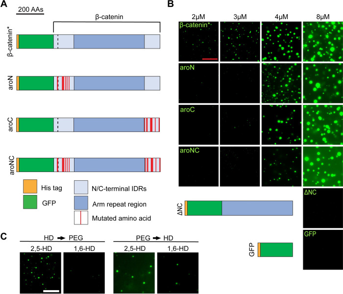
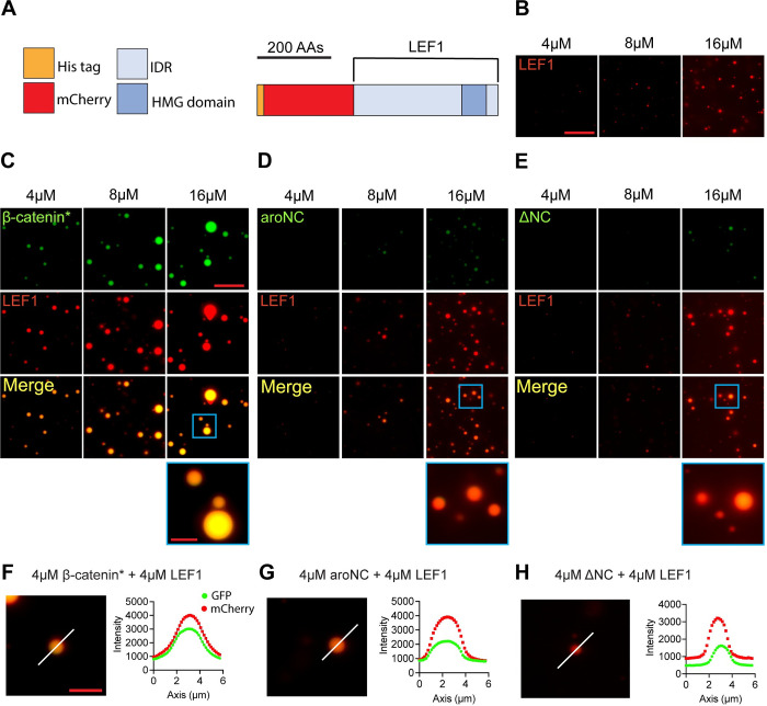
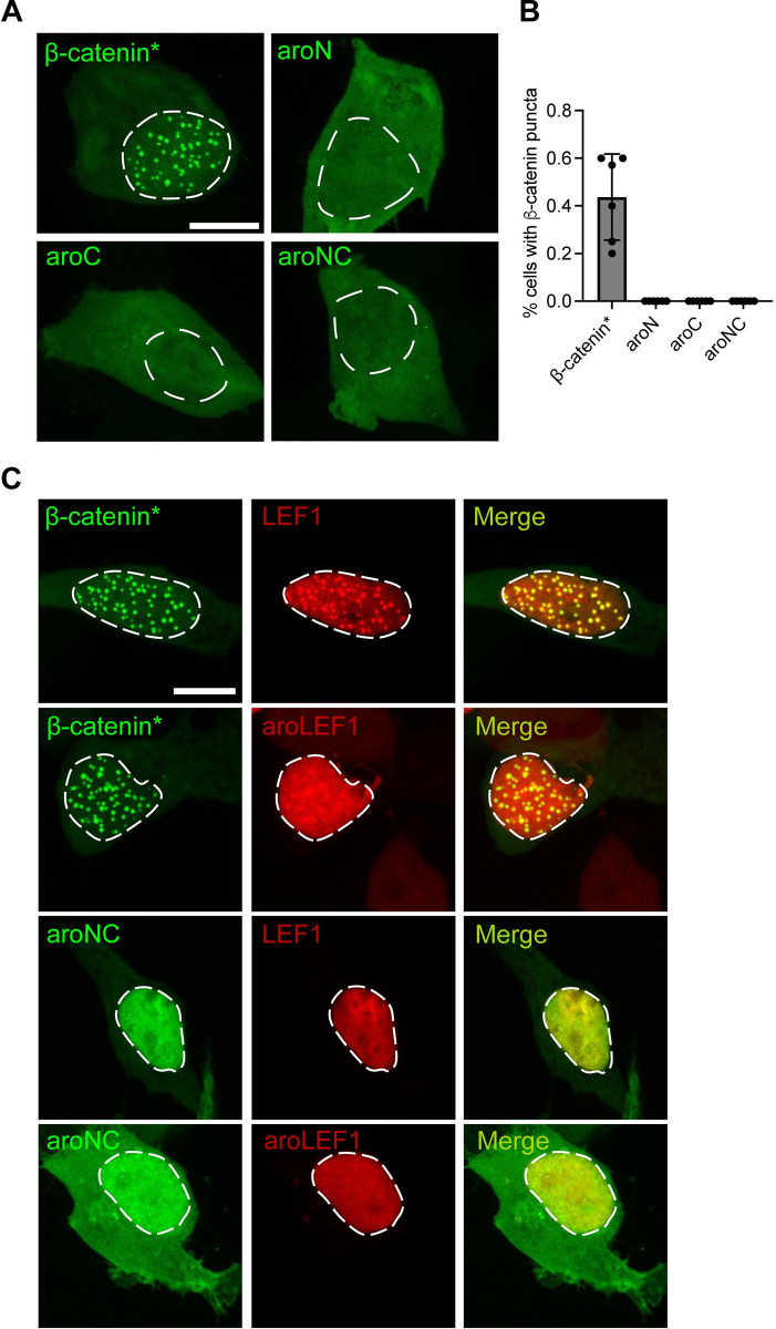
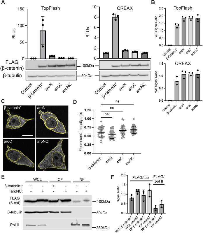
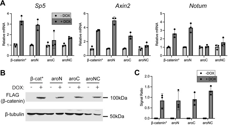
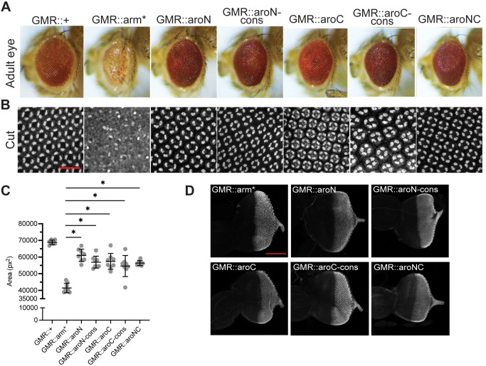
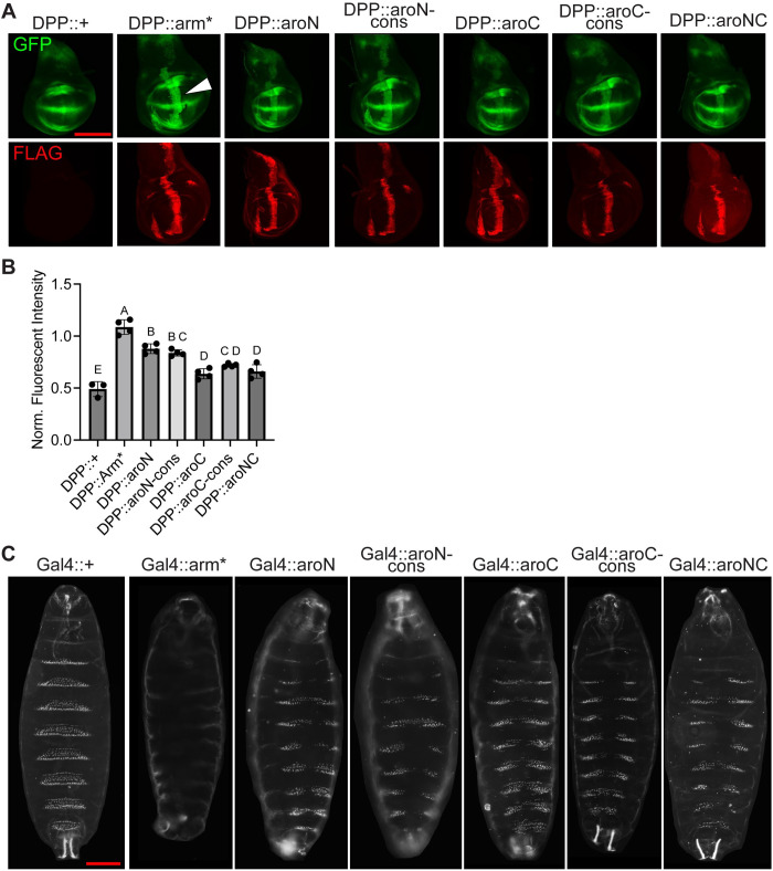
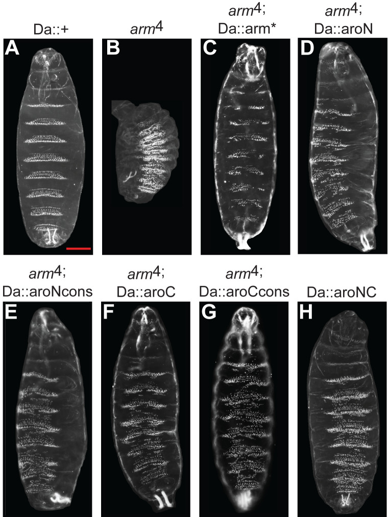
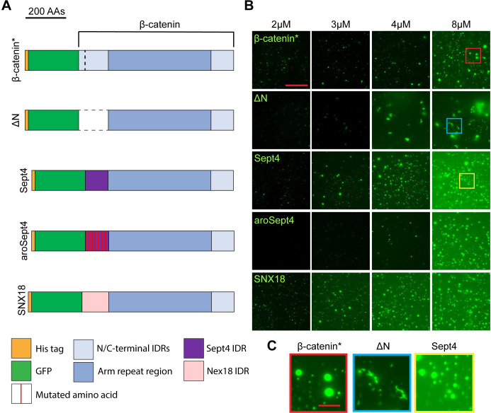
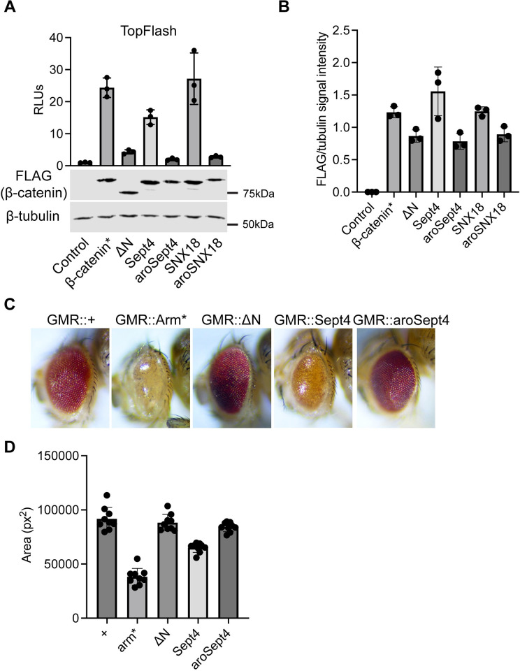
The Wnt/β-catenin signaling pathway plays numerous essential roles in animal development and tissue/stem cell maintenance. The activation of genes regulated by Wnt/β-catenin signaling requires the nuclear accumulation of β-catenin, a transcriptional co-activator. β-catenin is recruited to many Wnt-regulated enhancers through direct binding to T-cell factor/lymphoid enhancer factor (TCF/LEF) family transcription factors. β-catenin has previously been reported to form phase-separated biomolecular condensates (BMCs), which was implicated as a component of β-catenin's mechanism of action. This function required aromatic amino acid residues in the intrinsically disordered regions (IDRs) at the N- and C-termini of the protein. In this report, we further explore a role for β-catenin BMCs in Wnt target gene regulation. We find that β-catenin BMCs are miscible with LEF1 BMCs in vitro and in cultured cells. We characterized a panel of β-catenin mutants with different combinations of aromatic residue mutations in human cell culture and Drosophila melanogaster. Our data support a model in which aromatic residues across both IDRs contribute to BMC formation and signaling activity. Although different Wnt targets have different sensitivities to loss of β-catenin's aromatic residues, the activation of every target examined was compromised by aromatic substitution. These mutants are not defective in nuclear import or co-immunoprecipitation with several β-catenin binding partners. In addition, residues in the N-terminal IDR with no previously known role in signaling are clearly required for the activation of various Wnt readouts. Consistent with this, deletion of the N-terminal IDR results in a loss of signaling activity, which can be rescued by the addition of heterologous IDRs enriched in aromatic residues. Overall, our work supports a model in which the ability of β-catenin to form biomolecular condensates in the nucleus is tightly linked to its function as a transcriptional co-regulator.


Conflict of interest statement

The authors have declared that no competing interests exist.

Figures

Fig 1
Fig 1. Aromatic amino acid residues in the terminal IDRs of β-catenin promote biomolecular condensate formation in vitro.
(A) Cartoon representation of the eGFP-β-catenin* protein, the aromatic mutant derivatives, and control constructs. β-catenin* contains one S33Y mutation (dashed black line) in the N-IDR. aroN has all 9 endogenous aromatic amino acids within the N-IDR mutated to alanines. aroC has all 10 endogenous aromatic amino acids within the C-IDR mutated to alanines. aroNC contains all 19 aromatic amino acid mutations. The aromatic mutant constructs contain the same S33Y mutation as β-catenin*. (B) Representative images from an in vitro droplet formation assay with the indicated mutants. Droplet assays were performed in 300 mM NaCl and 10% PEG-8000. Scale bar = 20 μm. (C) Representative images from an in vitro droplet formation assay testing eGFP-β-catenin* sensitivity to 1,6-hexanediol and 2,5-hexanediol (used as a control); 8 μm eGFP-β-catenin* protein was exposed to the hexanediols prior to PEG-8000 (HD -> PEG) or PEG-8000 prior to the hexanediols (PEG -> HD), and 10% hexanediol and 10% PEG-8000 was used. Scale bar = 20 μm. IDR, intrinsically disordered region.
Fig 2
Fig 2. The β-catenin terminal IDRs promote β-catenin incorporation into LEF1 condensates in vitro.
(A) Cartoon representation of the mCherry-LEF1 construct. (B) Representative images from an mCherry-LEF1 in vitro droplet formation assay. Equal amounts of mCherry-LEF1 and eGFP (used as a control) were present in each reaction. Assays were performed in 300 mM NaCl and 10% PEG-8000. Scale bar = 20 μm. (C–E) Representative images from heterotypic in vitro droplet formation assays. (C) Equal amounts of eGFP-β-catenin* and mCherry-LEF1, (D) eGFP-aroNC and mCherry-LEF1, and (E) eGFP- ΔNC and mCherry-LEF1 were mixed, resulting in total protein concentrations of 4 μm, 8 μm, and 16 μm. Reaction conditions are the same as panel B. Scale bar = 20 μm, inset scale bar = 5 μm. (F–H) Line plots showing fluorescent intensity across a droplet. (F) eGFP-β-catenin* + mCherry-LEF1, (G) eGFP-aroNC and mCherry-LEF1, and (H) eGFP-ΔNC and mCherry-LEF1. Increased fluorescent signal for both proteins across a line indicates co-localization and the white lines represent the plotted trace. Scale bar = 5 μm. Summary data displayed in Fig 2 can be found in S1 Data. IDR, intrinsically disordered region.
Fig 3
Fig 3. Aromatic amino acid residues within β-catenin and LEF1 promote biomolecular condensate formation in vivo.
(A) Representative confocal images of HEK293T β-catenin KO cells expressing β-catenin mutants. Cells were transiently transfected with constructs encoding the FLAG-eGFP-β-catenin mutants. Scale bar = 10 μm. (B) Quantification of the percentage of cells that exhibited puncta. Data are presented as mean ± SD (n = 3). (C) Representative confocal images of HEK293T β-catenin KO cells expressing the indicated β-catenin and LEF1 constructs. Cells were transiently transfected with constructs encoding the FLAG-eGFP-β-catenin mutants and the FLAG-mCherry-LEF1 mutants. Scale bar = 10 μm. Summary data displayed in Fig 3 can be found in S1 Data.
Fig 4
Fig 4. Aromatic residues within β-catenin’s terminal IDRs are required for reporter gene activation and not nuclear accumulation.
(A) TopFlash (left) or CREAX (right) luciferase reporter activity induced by β-catenin* or the aromatic mutant constructs in HEK293T cells. Cells were transfected with separate plasmids encoding the reporter genes and the FLAG-β-catenin mutant constructs or pcDNA3.1 as a negative control. Corresponding western blots show relative expression of the FLAG-β-catenin mutant constructs. (B) Quantification of western blots from 3 independent replicate experiments for A. Data presented as the ratio of FLAG signal intensity to β-tubulin signal intensity. (C) Representative IF images of HEK293T cells for the indicated FLAG-β-catenin mutants. Cells were transfected with plasmids encoding the FLAG-β-catenin mutant constructs. IF was performed 24 h after transfection and nuclei were stained with DAPI. The borders of the cell and nucleus are highlighted. Scale bar = 10 μm. (D) Quantification of IF showing no significant difference in nuclear localization between β-catenin* and aromatic mutants. Data is presented as a ratio of the fluorescent intensity within the nucleus to the fluorescent intensity outside the nucleus. (E) Western blot showing nuclear fractionation samples from HEK293T cells expressing FLAG-β-catenin* or FLAG-aroNC. (F) Quantification of western blots from 3 independent experiments. Data presented as the ratio of FLAG signal intensity to either β-tubulin or RNA pol II signal intensity. All data are presented as mean ± SD; p-values were calculated by one-way ANOVA followed by Dunnett’s test. ns = p > 0.05. Summary data displayed in Fig 4 can be found in S1 Data. IDR, intrinsically disordered region; IF, immunofluorescence.
Fig 5
Fig 5. Select Wnt target genes exhibit different sensitivities to β-catenin aromatic mutant constructs.
(A) qRT-PCR analysis of 3 Wnt target genes (Sp5, Axin2, and Notum) in HeLa cells that were stably transformed with DOX-inducible, β-catenin mutant expression vectors. Cells were treated with DOX for 24 h prior to harvesting. (B) Western blot analysis of DOX-treated HeLa cell lysate. Lysate samples correspond to the qPCR data. α-FLAG blot shows β-catenin expression. α-tubulin was used as a loading control. (C) Quantification of western blots from 3 independent experiments. Data presented as the ratio of FLAG signal intensity to tubulin. All data presented as mean ± SD (n = 3). Summary data displayed in Fig 5 can be found in S1 Data.
Fig 6
Fig 6. β-catenin/Arm activity in the adult Drosophila eye is attenuated by aromatic amino acid mutations within the terminal IDRs.
(A) Micrographs of adult Drosophila eyes containing P[GMR-Gal4] and various P[UAS-Arm] transgenes. (B) Representative images of pupal Drosophila eye tissue immunostained for the cone cell marker Cut. Stabilized Arm (Arm*) disrupts cone cell specification while aromatic mutants do not. Scale bar = 20 μm. (C) Quantification of adult Drosophila eye area. Data are presented as mean ± SD (n = 8); p-values were calculated by one-way ANOVA followed by Dunnett’s test. * = p < 0.05. (D) Expression of the various Arm constructs is constant during late larval eye development. Representative images of larval Drosophila eye antennal discs immunostained for FLAG, representing β-catenin mutant expression. Scale bar = 100 μm. Summary data displayed in Fig 6 can be found in S1 Data. IDR, intrinsically disordered region.
Fig 7
Fig 7. Aromatic β-catenin/Arm mutants exhibit different levels of activity in wing imaginal discs and embryonic epidermis.
(A) Representative images of late third instar wing imaginal discs showing expression of a synthetic Wnt GFP reporter combined with a P[Dpp-Gal4] driving expression of various P[UAS-Arm] transgenes. Discs were also immunostained with α-FLAG to detect expression of the various Arm mutants. Gal4 activity was restricted to 18 h before fixation using a Gal80ts transgene. Scale bar = 100 μm. (B) Quantification of the synthetic Wnt GFP reporter activity. Reporter activity driven by ectopic expression of Arm mutants was normalized to reporter activity driven by endogenously expressed Wg/Arm signaling. Letters above bars indicate statistical significance (p < 0.05, calculated by one-way ANOVA followed by Dunnett’s test). Bars with the same letter above them are not significantly different, bars with different letters are. (C) Representative darkfield images showing the ventral side of late embryonic Drosophila cuticles containing P[Da-Gal4], P[Arm-Gal4], and 2 copies of the various P[UAS-Arm] transgenes. Scale bar = 100 μm. Summary data displayed in Fig 7 can be found in S1 Data.
Fig 8
Fig 8. Aromatic β-catenin/Arm mutants partially rescues an arm loss-of-function allele.
(A) Ventral side of a cuticle from a Drosophila embryo containing the P[Da-Gal4] transgene. Phenotype is indistinguishable from wild-type. (B) Cuticle of an amorphic arm mutant (arm4), displaying a classic Wg loss-of-function phenotype. (C) Cuticle of an arm mutant embryo containing P[Da-Gal4] and P[UAS-Arm*]. This combination results in nearly complete rescue of the size, head and posterior and lawn of denticle arm phenotype with 100% penetrance. (D–H), Cuticles of arm mutants embryos containing P[Da-Gal4] and either P[UAS-aroN] (D), P[UAS-aroNcons] (E), P[UAS-aroC] (F), P[UAS-aroCcons] (G), or P[UAS-aroNC] (H). These embryos have rescued the size, head and posterior phenotypes, but not the ectopic denticles.
Fig 9
Fig 9. Heterologous IDRs rescue the in vitro droplet formation of an N-terminal β-catenin deletion mutant.
(A) Cartoon representation of the eGFP-β-catenin* protein, ΔN (amino acids 1–151 deleted), Sept4 (119 amino acids from the Septin4 IDR), aroSept4 (Sept4 IDR with 11 aromatic amino acid mutations), and SNX18 (139 amino acids from the SNX18 IDR). (B) Representative images from a concentration series in vitro droplet formation assay with the indicated mutants. Droplet assays were performed in 300 mM NaCl and 10% PEG-8000. Scale bar = 20 μm. (C) Insets from the highlighted regions of panel B. IDR, intrinsically disordered region.
Fig 10
Fig 10. Heterologous IDRs can rescue the activity of an N-terminal β-catenin deletion mutant.
(A) TopFlash luciferase reporter activity induced by β-catenin* or the aromatic mutant constructs in HEK293T β-catenin KO cells. Cells were transfected with separate plasmids encoding the reporter gene and the FLAG-β-catenin mutant constructs or pcDNA3.1 as a negative control. Corresponding western blots show expression of the FLAG-β-catenin mutant constructs. Data are plotted as mean ± SD (n = 3). (B) Quantification of western blots from 3 independent replicate experiments for (A). (C) Micrographs of adult Drosophila eyes containing the indicated transgenes. (D) Quantification of adult Drosophila eye area. Data are presented as mean ± SD (n = 8). Summary data displayed in Fig 10 can be found in S1 Data. IDR, intrinsically disordered region.

Similar articles

References

    1. Rim EY, Clevers H, Nusse R. The Wnt Pathway: From Signaling Mechanisms to Synthetic Modulators. Annu Rev Biochem. 2022;91:571–598. doi: 10.1146/annurev-biochem-040320-103615 - DOI - PubMed
    1. Archbold HC, Yang YX, Chen L, Cadigan KM. How do they do Wnt they do?: regulation of transcription by the Wnt/β-catenin pathway: Wnt/β-catenin transcriptional regulation. Acta Physiologica. 2012;204:74–109. doi: 10.1111/j.1748-1716.2011.02293.x - DOI - PubMed
    1. Mosimann C, Hausmann G, Basler K. β-Catenin hits chromatin: regulation of Wnt target gene activation. Nat Rev Mol Cell Biol. 2009;10:276–286. doi: 10.1038/nrm2654 - DOI - PubMed
    1. Pagella P, Söderholm S, Nordin A, Zambanini G, Ghezzi V, Jauregi-Miguel A, et al.. The time-resolved genomic impact of Wnt/β-catenin signaling. Cell Systems. 2023;14:563–581.e7. doi: 10.1016/j.cels.2023.06.004 - DOI - PubMed
    1. Nusse R, Clevers H. Wnt/β-Catenin Signaling, Disease, and Emerging Therapeutic Modalities. Cell. 2017;169:985–999. doi: 10.1016/j.cell.2017.05.016 - DOI - PubMed

MeSH terms

LinkOut - more resources