. 2024 Dec:50:102125.
doi: 10.1016/j.tranon.2024.102125. Epub 2024 Sep 23.

MicroRNA-376a-3p sensitizes CPT-11-resistant colorectal cancer by enhancing apoptosis and reversing the epithelial-to-mesenchymal transition (EMT) through the IGF1R/PI3K/AKT pathway

Affiliations

MicroRNA-376a-3p sensitizes CPT-11-resistant colorectal cancer by enhancing apoptosis and reversing the epithelial-to-mesenchymal transition (EMT) through the IGF1R/PI3K/AKT pathway

Chikondi Jassi et al. Transl Oncol. 2024 Dec.

Abstract

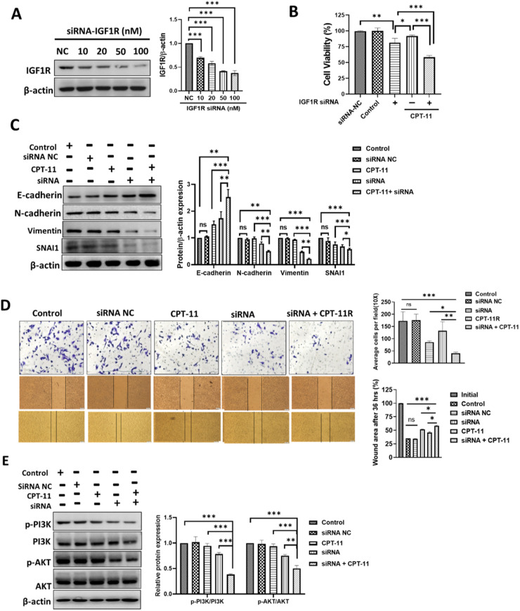
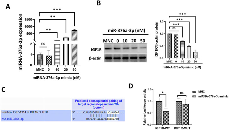
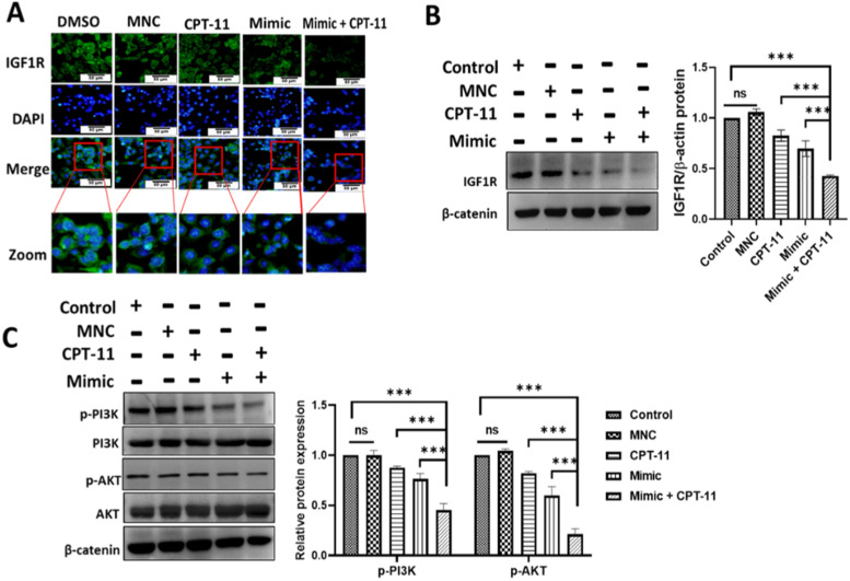
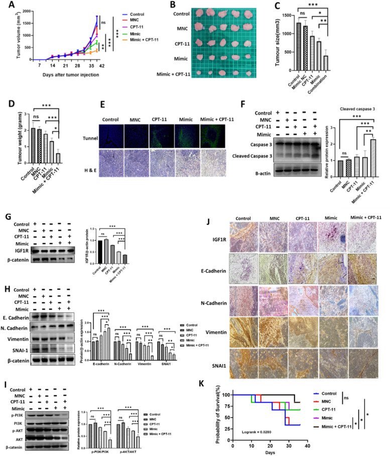
Colorectal cancer (CRC) remains the third most prevalent type of cancer worldwide contributing to an estimated 10 % of all cancer cases. CPT-11 is one of the first-line drugs for CRC treatment. Unfortunately, the development of drug resistance significantly exacerbates the adverse impact of CRC. Consequent tumor recurrences and metastasis, years after treatment are the frequently reported incidences. MicroRNAs (miRNA) are short non-coding RNA with the functionality of gene suppression. The insulin-like growth factor type 1 receptor (IGF1R) is a tyrosine kinase receptor frequently upregulated in cancers and is associated with cell survival and drug resistance. MiRNAs are frequently reported to be dysregulated in cancers including CRC. Evidence suggests that dysregulated miRNAs have direct consequences on the biological processes of their target genes. We previously demonstrated that miRNA-376a-3p is upregulated in CPT-11responsive, CRC cells upon treatment with CPT-11. We therefore aimed to investigate the involvement of miRNA-376a-3p in CPT-11 resistance and its probable association with IGF1R-mediated cancer cell survival. Our experimental approach used knockdown and overexpression experiments supplemented with western blot, RT-qPCR, flow cytometry, MTT, and migration assays to achieve our aim. Our data reveals the mechanism through which IGF1R and miRNA-376a-3p perpetrate and attenuate CPT-11 resistance respectively. MiRNA-376a-3p overexpression negatively regulated the IGF1R-induced cell survival, PI3K/AKT pathway, and reversed the epithelial-mesenchymal transition, hence sensitizing resistant cells to CPT-11. Our findings suggests that the miRNA-376a-3p/IGF1R axis holds promise as a potential target to sensitize CRC to CPT-11 in cases of drug resistance.

Keywords: CPT-11; CPT-11 resistance; Colorectal cancer; IGF1R; miRNA-376a-3p.


Conflict of interest statement

Declaration of competing interest The authors declare that they have no known competing financial interests or personal relationships that could have appeared to influence the work reported in this paper.

Figures

Fig. 1
Fig. 1
CPT-11 resistance is associated with epithelial-mesenchymal transition (EMT) and cell migration. The resistance of colorectal cancer cells to CPT-11 (CPT-11R) was confirmed using MTT assays, comparing them to parental (LoVo) cells (A-C). Topoisomerase I expression was evaluated to compare the response of these two cell types (D). CPT-11R cells exhibited morphological features associated with invasiveness and demonstrated higher migratory potential compared to parental cells (E-G). The expression levels of epithelial-to-mesenchymal transition (EMT) markers E-cadherin, N-cadherin, vimentin and SNAI1 were compared among untreated Caco2, LoVo, and CPT-11R cells (H). Additionally, the responsiveness of these cell lines to CPT-11 treatment was evaluated in the context of EMT markers (I). Finally, parental LoVo cells and CPT-11R cells were compared in their response to CPT-11 treatment in terms of migration, morphological changes, and apoptosis (J). Results are presented as means ± standard deviation from three independent experiments. Statistical significance was determined with the following annotations: ns (not significant). ns: not significant, p < 0.05 (*p < 0.05, ** <0.01, *** p < 0.001). CPT-11R: CPT-11 resistant cells established from LoVo cells, EMT: Epithelial-Mesenchymal Transition.
Fig. 2
Fig. 2
MiRNA-376a-3p is downregulated in CPT-11 resistant colorectal cancer cells. The downregulation of miRNA-376a-3p due to CPT-11 treatment in LoVo cells was confirmed by RT-qPCR (A). The expression of miRNA-376a-3p in CPT-11 resistant cells was also determined by RT-qPCR (B). Additionally, the effect of CPT-11 treatment on the expression of miRNA-376a-3p in CPT-11 resistant cells was investigated (C). Results are presented as means ± standard deviation from three independent experiments. Statistical significance was determined with the following annotations: ns (not significant). ns: not significant, p < 0.05 (*p < 0.05, ** <0.01, *** p < 0.001) CPT-11R: CPT-11 resistant cells established from LoVo cells.
Fig. 3
Fig. 3
IGF1R and its downstream PI3K/ AKT pathway are upregulated in CPT-11 resistant cells. Online bioinformatic tools, namely TargetScan, miRTarbase and miRTargetLink, were used to identify genes predicted to be targeted by miRNA-376a-3p (A). A total of sixteen genes were identified, overlapping across the three tools. Among, was IGF1R (B). The DAVID online bioinformatic platform was used to elucidate biological functions and pathways associated with these genes. Among them was PI3K/AKT pathway aligned with IGF1R (C). Before proceeding with the subsequent experiments, the expression of IGF1R in CPT-11R cells was evaluated by western blot analysis and immunofluorescence, comparing with that of LoVo cells as well as another CRC cell line Caco2 (D-E). The expression of downstream survival regulated by IGF1R, the PI3K/AKT pathway, and its response to CPT-11 treatment in CPT-11R were evaluated in CPT-11R cells and compared to their LoVo cells (F). Results are presented as means ± standard deviation from three independent experiments. Statistical significance was determined with the following annotations: ns (not significant). ns: not significant p < 0.05 (*p < 0.05, ** <0.01, *** p < 0.001).
Fig. 4
Fig. 4
Downregulation of IGF1R sensitizes resistant cells to CPT-11. To explore the role of IGF1R in CPT-11R cells, IGF1R was knocked down using siRNA. Knockdown of IGF1R was confirmed by western blot (A). Subsequently, the impact of IGF1R knockdown on cell viability and EMT and sensitization to CPT-11 was assessed by MTT and western blot respectively (B-C). Additionally, transwell and wound healing assay were performed to determine the effect of IGF1R downregulation on response to CPT-11(D). Furthermore, IGF1R downstream, PI3K/AKT signaling pathway was assessed for its response to IGF1R knockdown (E). Results are presented as means ± standard deviation from three independent experiments. Statistical significance was determined with the following annotations: ns (not significant). ns: not significant, p < 0.05 (*p < 0.05, ** <0.01, *** p < 0.001).
Fig. 5
Fig. 5
MiRNA-376a-3p downregulates IGF1R in CPT-11 resistant LoVo cells. Cells were transfected to overexpress MicroRNA-376a-3p. Transfection efficiency was determined by RT-qPCR (A). The effect of miRNA-376a-3p mimic on IGF1R expression was assessed by western blot (B). To confirm the regulatory effect of miRNA-376a-3p on IGF1R, the TargetScan bioinformatics tool was utilized to identify the predicted target region of miRNA-376a-3p on the 3′ UTR of IGF1R (C). Subsequently, a luciferase activity assay was performed to validate this targeting (D). Results are presented as means ± standard deviation from three independent experiments. Statistical significance was determined with the following annotations: ns (not significant). ns: not significant, p < 0.05 (*p < 0.05, ** <0.01, *** p < 0.001). MNC: Mimic negative control, WT: Wild-type, MUT: Mutant-type.
Fig. 6
Fig. 6
MiR-376a-3p sensitizes CPT-11 resistant cells to CPT-11. Different concentrations of CPT-11 were co-administered with a miRNA-376a-3p mimic, along with respective controls, to assess the sensitizing impact of miRNA-376a-3p on cell viability using MTT assay (A). Co-treatment of miRNA-376a-3p with CPT-11 resulted in greater enhancement of the expression of cleaved PARP and caspase 3 compared to individual treatments and controls, indicating an increase in apoptosis (B). Apoptosis-enhancing effect of miRNA-376a-3p in combination with CPT-11 was further confirmed with TUNEL assay (C). The effect of miRNA-376a-3p mimic on EMT was assessesd by western blot (D). The inhibition of cell migration was significantly enhanced by the combination of miRNA-376a-3p and CPT-11 in comparison to both individual treatments and the control group. This effect was assessed using transwell and wound healing migration assays (E-F). Results are presented as means ± standard deviation from three independent experiments. Statistical significance was determined with the following annotations: ns (not significant). ns: not significant, p < 0.05 (*p < 0.05, ** <0.01, *** p < 0.001). MNC: Mimic negative control.
Fig. 7
Fig. 7
MicroRNA-376a-3p sensitizes CPT-11 resistant cells to CPT-11 through PI3K/AKT pathway via IGF1R downregulation. To determine the mechanism by which miRNA-376a-3p sensitizes CPT-11R cells, we transfected cells with miRNA-376a-mimic, CPT-11, their combination and the respective controls, and assessed the expression of IGF1R using both immunocytochemistry and western blot (A-B). Subsequently, we examined the expression of the downstream pathway of IGF1R, the PI3K/AKT pathway, through western blot analysis (C). Results are shown as means ± standard deviation from three independent experiments. ns: not significant, p < 0.05 (*p < 0.05, ** <0.01, *** p < 0.001). MNC: Mimic negative control.
Fig. 8
Fig. 8
MiRNA-376a-3p improved antitumor efficacy against murine CPT-11 resistant colorectal cancer xenograft in BALB/c nude mice. CPT-11 resistant tumors were induced by intracutaneously injecting CPT-11 resistant cells into the flanks of mice. Tumor growth was closely monitored both before and throughout the six-week treatment period (A). At the conclusion of the study, mice were humanely euthanized, and tumors were excised. Tumor sizes and weights were then measured and compared against controls (B-D). Subsequently, tumors were embedded in paraffin sections, sliced, and mounted on microscope slides for staining. TUNEL and H & E staining techniques were employed (E). Proteins were extracted from the tumors, and Western blot analysis was conducted to evaluate apoptotic marker, caspase 3 (F), IGF1R (G), EMT markers (H), and the expression of the PI3K/AKT pathway (I). Additionally, paraffin-embedded slides were utilized for immunohistochemistry to assess the expression of IGF1R, E-cadherin, N-cadherin,vimentin and SNAI1 (J). The survival probability of the mice involved in the study was predicted using Kaplan Meier survival curves (K). Results are presented as means ± standard deviation from three independent experiments. "ns" denotes non-significance, with statistical significance set at p < 0.05 (*p < 0.05, ** <0.01, *** p < 0.001). MNC stands for Mimic Negative Control.

References

    1. Sung H., et al. Global cancer statistics 2020: GLOBOCAN estimates of incidence and mortality worldwide for 36 cancers in 185 Countries. CA Cancer J. Clin. 2021;71(3):209–249. - PubMed
    1. Biller L.H., Schrag D. Diagnosis and treatment of metastatic colorectal cancer: a review. JAMA. 2021;325(7):669–685. - PubMed
    1. Makondi P.T., et al. Prediction of novel target genes and pathways involved in irinotecan-resistant colorectal cancer. PLoS ONE. 2017;12(7) - PMC - PubMed
    1. de Man F.M., et al. Individualization of Irinotecan Treatment: a Review of Pharmacokinetics, Pharmacodynamics, and Pharmacogenetics. Clin. Pharmacokinet. 2018;57(10):1229–1254. - PMC - PubMed
    1. Thomas A., Pommier Y. Targeting topoisomerase I in the era of precision medicine. Clin. Cancer Res. 2019;25(22):6581–6589. - PMC - PubMed

LinkOut - more resources