. 2024 Nov:77:103358.
doi: 10.1016/j.redox.2024.103358. Epub 2024 Sep 18.

Overexpression of NUDT16L1 sustains proper function of mitochondria and leads to ferroptosis insensitivity in colorectal cancer

Affiliations

Overexpression of NUDT16L1 sustains proper function of mitochondria and leads to ferroptosis insensitivity in colorectal cancer

Yi-Syuan Lin et al. Redox Biol. 2024 Nov.

Abstract

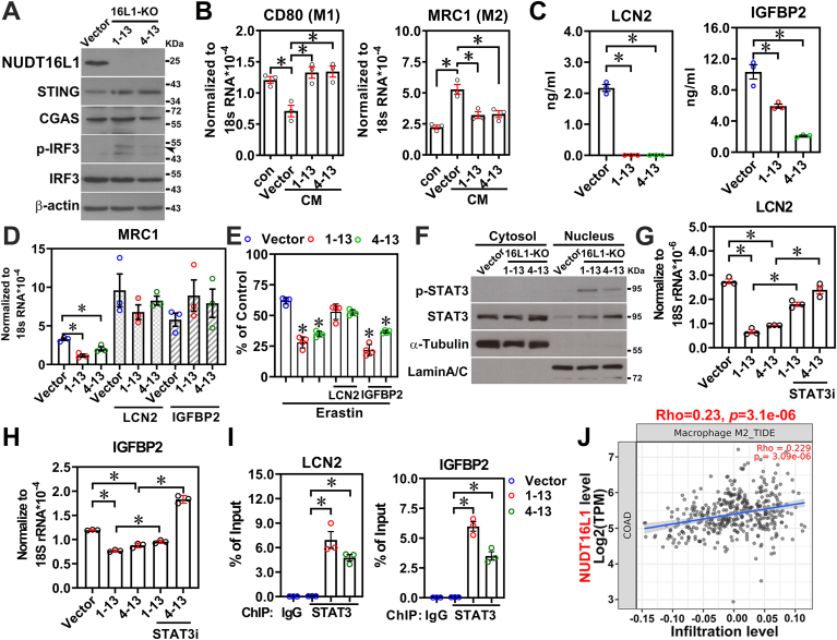
Cancer research is continuously exploring new avenues to improve treatments, and ferroptosis induction has emerged as a promising approach. However, the lack of comprehensive analysis of the ferroptosis sensitivity in different cancer types has limited its clinical application. Moreover, identifying the key regulator that influences the ferroptosis sensitivity during cancer progression remains a major challenge. In this study, we shed light on the role of ferroptosis in colorectal cancer and identified a novel ferroptosis repressor, NUDT16L1, that contributes to the ferroptosis insensitivity in this cancer type. Mechanistically, NUDT16L1 promotes ferroptosis insensitivity in colon cancer by enhancing the expression of key ferroptosis repressor and mitochondrial genes through direct binding to NAD-capped RNAs and the indirect action of MALAT1. Our findings also reveal that NUDT16L1 localizes to the mitochondria to maintain its proper function by preventing mitochondrial DNA leakage after treatment of ferroptosis inducer in colon cancer cells. Importantly, our orthotopic injection and Nudt16l1 transgenic mouse models of colon cancer demonstrated the critical role of NUDT16L1 in promoting tumor growth. Moreover, clinical specimens revealed that NUDT16L1 was overexpressed in colorectal cancer, indicating its potential as a therapeutic target. Finally, our study shows the therapeutic potential of a NUDT16L1 inhibitor in vitro, in vivo and ex vivo. Taken together, these findings provide new insights into the crucial role of NUDT16L1 in colorectal cancer and highlight its potential as a promising therapeutic target.

Keywords: Colon cancer; Ferroptosis insensitivity; Mitochondrial DNA leakage; Mitochondrial function; NUDT16L1; Tumor growth; metabolite-cap, MALAT1 lncRNA.


Conflict of interest statement

Declaration of competing interest The authors declare that they have no known competing financial interests or personal relationships that could have appeared to influence the work reported in this paper.

Figures

Fig. 1
Fig. 1
NUDT16L1 was a novel ferroptosis repressor causing ferroptosis insensitivity in colorectal cancer. (A) The effects of GPX4 knockout on cell survival in different types of cancer cell lines were downloaded from DepMap (https://depmap.org/portal/). Dependency scores of GPX4 in different cancer cell lines from the same type of cancer were averaged to present (Gastric: n = 14; Esophageal: n = 26; Colon: n = 30; Lung: n = 40; Pancreatic: n = 23; Head and Neck: n = 33; Ovarian: n = 29; Neuroblastoma: n = 17; Myeloma: n = 5; Breast: n = 25; Prostate: n = 3; Brain: n = 33; Bone: n = 13). Negative score indicated poor cell survival phenomenon. (B) The cell viability of different cancer cell types was measured by trypan blue exclusion assay after different doses of ferroptosis inducers, RSL3 (0.25 μM and 1 μM) treatment for 24 h (n = 4). (C) The Heatmap showed the expression levels of GPX4 and members of NUDT family in TCGA COAD dataset (n = 287) and those results were performed Pearson correlation analysis. (D) The correlation analysis between gene expression level of NUDT16L1 and drug sensitivity was analyzed from Cancer Therapeutics Response Portal (https://portals.broadinstitute.org/ctrp/). Positive correlation indicated that the higher gene expression was associated drug insensitivity. (E) Total glutathione in NUDT16L1-KO HCT116 and its control cells was measured by Glutathione Colorimetric Detection Kit (n = 4). (F) The accumulation of ROS in NUDT16L1-KO HCT116 and its control cells was detected by staining CellROX Dye and the image was quantified by Image J (n = 3). (G) NUDT16L1-KO HCT116 and its control cells were sent to perform lipidomic analysis. Lipids were categorized into saturated fatty acids (SFA), monounsaturated fatty acids (MUFA), and polyunsaturated fatty acids (PUFA) based on the number of double bonds. The expression levels of these lipid species were then visualized as heatmaps within each group (SFA, MUFA, and PUFA), with no additional normalization applied to the lipidomic data presented. The number of Y axis as indicated the number of double bonds. (H, I) Mitochondrial morphology in NUDT16L1-KO HCT116 and its control cells was analyzed by transmission electron microscopy (TEM). Mitochondria was annotated as Mito (H). Mitochondria size was measured by Image J (I). (J) The accumulation of lipid peroxidation in NUDT16L1-KO and its-restored HCT116 cells was detected by staining BODIPY 581/591C11 dye. (K) Those images from (J) were quantified by Image J (n = 3). (L) Cell viabilities were measured by trypan blue exclusion assay in control and NUDT16L1-KO cells with/without different doses of RSL3 treatment for 24 h (n = 4). Results were shown as percentage of treatment control. (M) The accumulation of ROS in NUDT16L1-KO and its-restored HCT116 cells was detected by staining DCFDA dye and quantified by flow cytometry (n = 4). (N) Cell viabilities were assessed using the trypan blue exclusion assay in both control and NUDT16L1-KO cells, with or without the reintroduction of exogenous NUDT16L1 (16L1OE), following 24-h RSL3 treatment (n = 4). Results were shown as percentage of treatment control. (O) NUDT16L1-KO HCT116 cells were treated with RSL3 (1 μM) and combined with different types of inhibitors for 24 h (n = 4). Cell viabilities were measured after treatments. Fer1: Ferrostatin1; Tro: Trolox; DFO: Deferoxamine; ZVAD: Z-VAD-FMK, a pan-caspase inhibitor; BA1: Bafilomycin A1, an autophagy inhibitor. All the cell viabilities were measured by using the trypan blue exclusion assay. (For interpretation of the references to color in this figure legend, the reader is referred to the Web version of this article.)
Fig. 2
Fig. 2
NUDT16L1 positively regulated several crucial genes to repress ferroptosis via the function of MALAT1 and contributed to ferroptosis insensitivity in colon cancer cells. (A) RNA-seq results of NUDTL16L1 knockdown were analyzed the features of the upregulated-genes under erastin treatment via gene set enrichment analysis (GSEA). (B) The transcripts of GPX4, SLC3A2, SLC7A11, MALAT1 and MT-CO1 were interacted with NUDT16L1 by RIP assay from iNUDT16L1-FLAG HCT116 cells with or without doxycycline (1 μg/ml) induction for 24 h by qRT-PCR (n = 3). % of input: denotes that the binding signals of IgG and NUDT16L1 derived from transcripts are normalized against the input control, which comprises samples without undergoing RNA-immunoprecipitation (C) GPX4, SLC3A2, SLC7A11, MALAT1 and MT-CO1 expression levels were measured in NUDT16L1-KO and its control HCT116 cells by using qRT-PCR (n = 3). (D) Various RNA types, including uncapped, m7G-capped, and NAD-capped RNA, were used to probe NUDT16L1(16L1) recombinant protein via RNA Pull-Down Assay combined with Western Blot. (E) NAD-capped mitochondrial RNA (SLC7A11, MT-CO1, MT-CYB, and MT-ND1) levels in NUDT16L1-KO and control HCT116 cells were measured using the ONE-seq technique combined with qRT-PCR (n = 3). (F) CRISPR activation (CRISPRa) technique was applied to establish stable restoration of MALAT1 in NUDT16L1-KO HCT116 cells. Expression levels of GPX4, SLC3A2, and MALAT1 were measured in NUDT16L1-KO HCT116 cells with or without MALAT1 restoration by qRT-PCR (n = 3). (G) Lipid peroxidation in NUDT16L1-KO HCT116 cells with or without MALAT1 restoration was detected by staining BODIPY 581/591C11 dye and those images were quantified by Image J (n = 3). (H) NUDT16L1-KO HCT116 cells with or without MALAT1 restoration were treated with vehicle, RSL3 (0.25 and 1 μM) (Left panel) or erastin (2.5 and 10 μM) (right panel) for 24 h and cell viabilities were measured by counting cell numbers. (I) Expression levels of MALAT1, GPX4, SLC3A2 and NUDT16L1 in a large cohort of human colorectal cancer specimen (E-MTAB-990, n = 688) were used to perform Pearson correlation analysis.
Fig. 3
Fig. 3
NUDT16L1 was also located in the mitochondria to maintain its proper function by inhibition of mPTP activity to prevent mtDNA leakage into cytosol in colon cancer cells. (A) NUDT16L1 expression in the mitochondrial, cytosol and nuclear fractions of HCT116 was determined by Western blot. VDAC, α-Tubulin and lamin A/C were served as mitochondrial, cytosol and nuclear marker, respectively. (B) The cellular localization of NUDT16L1 in HCT116 cell was determined by immunogold staining combined with transmission electron microscopy (TEM). Red dashed line was indicated the position of mitochondria (Mito). (C) The function of mitochondria in HCT116 cells with NUDT16L1-WT, -KO and its restoration were measured by Seahorse XFe Analyzer (n = 4). (D) The mitochondrial ROS was measured in HCT116 cells with NUDT16L1-WT, -KO and its restoration by staining with MitoSOX, a mitochondrial superoxide indicator. Those results were quantified by flow cytometry (n = 4). (E) The mitochondrial membrane potential was measured in HCT116 cells with NUDT16L1-WT, -KO and its restoration by staining with TMRM, a mitochondrial membrane potential indicator. Those results were quantified by flow cytometry (n = 4). (F) The Mitochondria mass was measured in NUDT16L1-KO, its restoration and control HCT116 cells by staining with MitoTracker dye and quantified by flow cytometry (n = 4). (G) The accumulation of lipid peroxidation in HCT116 cells harboring NUDT16L1-WT, -KO, and their restored wild-type (WT) and mitochondrial-specific (MTS) forms was detected by BODIPY 581/591C11, and the fluorescence images were captured by fluorescence microscopy and quantified by Image J (n = 3). (H) Cell viabilities were measured by trypan blue exclusion assay in HCT116 cells harboring NUDT16L1-WT, -KO, and their restored wild-type (WT) and mitochondrial-specific (MTS) forms under treatments of ferroptosis inducers, RSL3 (1 μM) for 24 h (n = 4). Results were shown as the percentage of treatment control. (I) The expression level of MT-CO1, a mitochondrial gene, was quantified in cytosolic DNA extracts from HCT116 cells harboring NUDT16L1-WT, -KO, and their restored wild-type (WT) and mitochondrial-specific (MTS) forms using qPCR. Quantification was normalized to the level of 18s rDNA in genomic DNA (n = 3). (J) Mitochondrial permeability transition pore (mPTP) activities were determined in NUDT16L1-KO and its control HCT116 cells by mitochondrial permeability transition pore assay kit and quantified by flow cytometry (n = 3). (K) NUDT16L1-KO and control HCT116 cells were treated with Trolox (100 μM) for 24 h, and mPTP activity was measured by using a mPTP assay kit and quantified by flow cytometry (n = 3). (L) HCT116 cells were treated with Bz-423, a mPTP activator, (10 μM) for 24 h and mtDNA leakage level (MT-CO1) were determined by qPCR. Results were normalized to 18s rDNA (n = 3). (M) Gene in mtDNA was measured in the cytosolic DNA extractions of NUDT16L1-KO and its control HCT116 cells received treatment with or without TRO19622 (5 μM), a mitochondrial permeability transition pore (mPTP) inhibitor, for 24 h (n = 3) by qPCR and normalized to the level of 18s rDNA in genomic DNA (n = 3). (N, O) Cell viabilities were measured by trypan blue exclusion assay in two different clones of NUDT16L1-KO HCT116 cells, 1–13 (N) and 4–13 (O), co-treated with ferroptosis inducers, RSL3 (0.25 μM and 1 μM) and erastin (2.5 μM and 10 μM), and mPTP inhibitors, TRO19622 (5 μM) and cyclosporin A (10 μM), for 24 h (n = 4). (For interpretation of the references to color in this figure legend, the reader is referred to the Web version of this article.)
Fig. 4
Fig. 4
NUDT16L1 repressed CGAS-STING-STAT3 signaling pathway to promote M2 macrophage polarization by increases of LCN2 and IGFBP2 and decrease of MIF secretions in colon cancer cells. (A) Components of cGAS-STING signaling pathway including STING, cGAS, phosphorylation of-IRF3 (p-IRF3) and its total form were detected in NUDT16L1-KO and its control HCT116 cells by Western blot. (B) PMA-activated U937 cells were incubated with the conditioned media from NUDT16L1-KO and its control HCT116 cells for 24 h and expression levels of CD80 (left panel) and MRC1 (right panel) were measured by qRT-PCR (n = 3). (C) The concentrations of LCN2 and IGFBP2 in the conditioned media of NUDT16L1-KO and its control HCT116 cells were individually measured by ELISA (n = 3). (D) PMA-activated U937 cells were co-incubated with the conditioned media from NUDT16L1-KO and its control HCT116 cells and LCN2 (2 ng/ml) or IGFBP2 (10 ng/ml) for 24 h. MRC1 expression was measured by qRT-PCR (n = 3). (E) NUDT16L1-KO and its control HCT116 cells were co-treated with erastin (10 μM) and LCN2 (2 ng/ml) or IGFBP2 (10 ng/ml) for 24 h to determine cell viability by counting cell numbers (n = 4). (F) Phosphorylation of STAT3 (p-STAT3) and its total form were analyzed in the nuclear and cytosolic fractionations from NUDT16L1-KO and its control HCT116 cells by Western blot. (G) NUDT16L1-KO and its control HCT116 cells were treated with APTSTAT3-9R (10 μM) for 24 h to determine the expression levels of LCN2 by qRT-PCR (n = 3). (H) NUDT16L1-KO and its control HCT116 cells were treated with APTSTAT3-9R (10 μM) for 24 h to determine the expression levels of IGFBP2 by qRT-PCR (n = 3). (I) Chromatin Immunoprecipitation (ChIP) by STAT3 or control IgG antibody was applied to verify its binding ability on gene locus of LCN2 (left panel) or IGFBP2 (right panel) in NUDT16L1-KO and its control HCT116 cells by qRT-PCR (n = 3). (J) Correlation analysis between NUDT16L1 expression and M2 macrophage infiltration in TCGA COAD dataset was analyzed by TIMER2.0 database (http://timer.cistrome.org/).
Fig. 5
Fig. 5
Overexpression of NUDT16L1 promoted the cancer development in the animal models of colon cancer. (A) Cell proliferation of NUDT16L1-KO and its control HCT116 cells was measured by counting cell numbers via trypan blue exclusion assay (n = 3). (B) NUDT16L1-KO and its control HCT116 cells were used to perform orthotopic injection into the caecum of NOD-SCID mice for one month. Representative images of tumor (B) and quantification results of their tumor weight (C) were shown (n = 6). (C) Quantification results of their tumor weight from (B) were shown (n = 6). (D) The expression level of Ym1, a M2 macrophage marker was shown in xenograft tumors of NUDT16L1-KO and its control HCT116 cells by using IHC staining method. (E) C57BL/6J-Nudt16l1-KO and its control mice were received AOM/DSS treatment to induce the colorectal carcinogenesis. Mouse colorectal tissues were performed Hematoxylin and Eosin (H&E) staining after sacrifice. The representative H&E images were shown. (F) Tumor areas from (E) were quantified by Image J (WT, n = 5; 16l1-KO, n = 4). (G) Vil1(villin)-Cre and Vil1-Cre cross with C57BL/6J-Nudt16l1-cKI mice were received AOM/DSS treatment to induce the colorectal carcinogenesis. Mouse colorectal tissues were performed Hematoxylin and Eosin (H&E) staining after sacrifice. The representative H&E images were shown. (H) Tumor areas from (G) were quantified by Image J (Vil-Cre, n = 5; Vil-Cre/16l1-CKI, n = 4). (For interpretation of the references to color in this figure legend, the reader is referred to the Web version of this article.)
Fig. 6
Fig. 6
NUDT16L1 was overexpressed in the clinical specimens of colorectal cancer. (A) NUDT16L1 expression was analyzed in GSE37812 from GEO dataset containing normal and colon cancer specimens (Normal, n = 88; cancer, n = 84). (B) NUDT16L1 expression was analyzed in TCGA COAD dataset (Normal, n = 301; COAD, n = 287). (C) The expression levels of NUDT16L1 in the epithelial cells of clinical colon cancer specimen was analyzed in the single cell RNA-seq dataset from GSE132465 (Normal, n = 344; Cancer, n = 11972). (D) The expression levels of GPX4 and SLC3A2 in the epithelial cells of clinical colon cancer specimen was analyzed in the single cell RNA-seq dataset from GSE132465 (Normal, n = 344; Cancer, n = 11972). (E) The expression levels of LCN2 and IGFBP2 in the epithelial cells of clinical colon cancer specimen was analyzed in the single cell RNA-seq dataset from GSE132465 (Normal, n = 344; Cancer, n = 11972). (F) MALAT1 expression in the clinical specimens of colorectal cancer was analyzed by GEO dataset (GSE37812) (Normal, n = 88; Cancer, n = 84) dataset. (G) NUDT16L1 expression was measured in our paired normal and colorectal cancer specimens by qRT-PCR (n = 49). (H) NUDT16L1 expression was measured in our paired normal and colorectal cancer specimens by IHC staining. Representative images of NUDT16L1 staining results containing normal (upper panel), and cancer specimens (lower panel) were shown. (I) NUDT16L1 IHC staining results from (H) were quantified by HistoQuest software (Normal, n = 328; Cancer, n = 384). (J) The expression levels of NUDT16L1 in the clinical specimens of colorectal cancer were used to perform its correlation with progression free survival in TCGA COAD dataset (High expression, n = 251; Low expression, n = 245).
Fig. 7
Fig. 7
Specific inhibitor of NUDT16L1 promoted ferroptosis and inhibited colon tumor growth. (A) The expression levels of NUDT16L1 and NUDT16 were analyzed in HCT116 cells treated with different doses of sophoranone for 48 h by Western blot (upper panel) and quantification results of NUDT16L1 were shown (lower panel) (n = 3). (B) Cell viability was measured by counting cell numbers in HCT116 cells treated with vehicle or different doses of sophoranone for different time points (n = 4). (C) Cell viabilities were measured by trypan blue exclusion assay in control and NUDT16L1-KO cells received different doses of sophoranone (0, 1.25, 2.5, 5, 10, and 20 μM) treatment for 24 h. (D)Lipid Peroxidation was measured in HCT116 cells treated with different doses of sophoranone by staining with BODIPY 581/591C11 and those images were quantified by Image J (n = 3). (E) Cellular ROS was measured in HCT116 cells received different doses of sophoranone treatment for 24 h by using DCFDA dye for flow cytometry (n = 4). (F) The function of mitochondria in HCT116 cells received different doses of sophoranone (0, 5, 10, 20 μM) treatment for 24 h were measured by Seahorse XFe Analyzer (n = 4). (G) Mitochondrial mass was analyzed in HCT116 cells received different doses of sophoranone (0, 5, 10, 20 μM) treatment for 24 h by staining with MitoTracker and quantified by flow cytometry (n = 4). (H) ATP levels were measured by StayBrite™ Highly Stable ATP bioluminescence assay kit in HCT116 cells received different doses of sophoranone for 24 h (n = 3). (I) Expression level of cytosolic mtDNA such as MT-CO1was measured by qPCR in HCT116 cells received different doses of sophoranone for 24 h (n = 3). (J) Phosphorylation of STAT3 (p-STAT3) and its total form were measured in HCT116 cells treated with different doses of sophoranone for 24 h by using Western blot. (K) LCN2 expression was detected in in HCT116 cells treated with different doses of sophoranone for 24 h by using qRT-PCR. (L) HCT116 cells were co-treated with different does of sophoranone and RSL3 (0.25 μM) or erastin (2.5 μM) for 24 h. Cell viability was measured by directly counting cell numbers and results were normalized to vehicle control (n = 4). (M, N) Human CRC organoids were individually treated with erastin (2.5 μM), sophoranone (sopho, 5 μM) or 5-FU (50 μM) as a positive control, and combination treatment of erastin and sophoranone for 3 days. The viability (M) and cell death (N) of CRC organoid were respectively determined by alamarblue assay and acridine orange (AO, lived cells) and propidium iodide (PI, dead cells) staining. (For interpretation of the references to color in this figure legend, the reader is referred to the Web version of this article.)
Fig. 8
Fig. 8
A cartoon briefly illustrated the critical role of NUDT16L1 in promoting both tumor growth and the development of ferroptosis insensitivity in colorectal cancer. Figure is created by BioRender.com.

References

    1. Dixon S.J., Lemberg K.M., Lamprecht M.R., Skouta R., Zaitsev E.M., Gleason C.E., et al. Ferroptosis: an iron-dependent form of nonapoptotic cell death. Cell. 2012;149:1060–1072. - PMC - PubMed
    1. Chen X., Kang R., Kroemer G., Tang D. Broadening horizons: the role of ferroptosis in cancer. Nat. Rev. Clin. Oncol. 2021 - PubMed
    1. Jiang X., Stockwell B.R., Conrad M. Ferroptosis: mechanisms, biology and role in disease. Nat. Rev. Mol. Cell Biol. 2021 - PMC - PubMed
    1. Badgley M.A., Kremer D.M., Maurer H.C., DelGiorno K.E., Lee H.J., Purohit V., et al. Cysteine depletion induces pancreatic tumor ferroptosis in mice. Science. 2020;368:85–89. - PMC - PubMed
    1. Ghoochani A., Hsu E.C., Aslan M., Rice M.A., Nguyen H.M., Brooks J.D., et al. Ferroptosis inducers are a novel therapeutic approach for advanced prostate cancer. Cancer Res. 2021;81:1583–1594. - PMC - PubMed

Publication types

Substances