. 2024 Oct 15;5(10):101744.
doi: 10.1016/j.xcrm.2024.101744. Epub 2024 Sep 23.

Pig-to-human kidney xenotransplants using genetically modified minipigs

Affiliations

Pig-to-human kidney xenotransplants using genetically modified minipigs

Yi Wang et al. Cell Rep Med. .

Abstract

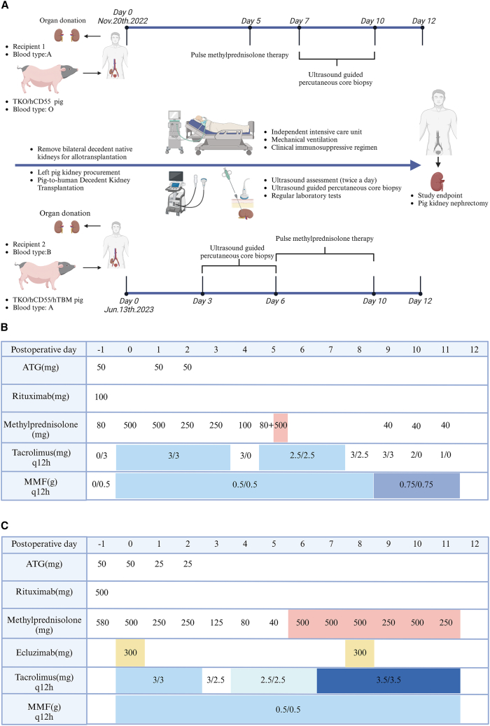
This study develops an observational model to assess kidney function recovery and xenogeneic immune responses in kidney xenotransplants, focusing on gene editing and immunosuppression. Two brain-dead patients undergo single kidney xenotransplantation, with kidneys donated by minipigs genetically modified to include triple-gene knockouts (GGTA1, β4GalNT2, CMAH) and human gene transfers (hCD55 or hCD55/hTBM). Renal xenograft functions are fully restored; however, immunosuppression without CD40-CD154 pathway blockade is ineffective in preventing acute rejection by day 12. This rejection manifests as both T cell-mediated rejection and antibody-mediated rejection (AMR), confirmed by natural killer (NK) cell and macrophage infiltration in sequential xenograft biopsies. Despite donor pigs being pathogen free before transplantation, xenografts and recipient organs test positive for porcine cytomegalovirus/porcine roseolovirus (PCMV/PRV) by the end of the observation period, indicating reactivation and contributing to significant immunopathological changes. This study underscores the critical need for extended clinical observation and comprehensive evaluation using deceased human models to advance xenograft success.

Keywords: PCMV/PRV; acute xenograft rejection; brain-dead human decedent; genetically engineered pig; kidney transplantation; xenotransplantation.


Conflict of interest statement

Declaration of interests The authors declare no competing interests.

Figures

None
Graphical abstract
Figure 1
Figure 1
Research process and immunosuppression regimen (A) Two cases of porcine-to-human decedent kidney transplantation: research timeline and event summary. (B) Pharmacologic immunosuppression regimen of case 1. (C) Pharmacologic immunosuppression regimen of case 2.
Figure 2
Figure 2
Clinical outcomes and immunological monitoring of case 1 throughout the postoperative course (A) Gross findings from the xenograft: xenograft perfusion (A1), xenograft reperfusion (A2), xenograft at termination (A3), and cross-section of the xenograft (A4). (B) Changes in urine output, serum creatinine, platelets, hemoglobin, tacrolimus concentration, and lymphocyte subsets. (C) Changes in antibody binding (IgM and IgG) to donor pig PBMCs and CDC against the same PBMCs (Mix: the pooled sera of 20 healthy human volunteers were used as control sera).
Figure 3
Figure 3
Clinical outcomes and immunological monitoring of case 2 throughout the postoperative course (A) Gross findings from the xenograft: xenograft perfusion (A1), xenograft reperfusion (A2), xenograft at termination (A3), and cross-section of the xenograft (A4). (B) Changes in urine output, serum creatinine, platelets, hemoglobin, tacrolimus concentration, and lymphocyte subsets. (C) Changes in antibody binding (IgM and IgG) to donor pig PBMCs and CDC against the same PBMCs (Mix: the pooled sera of 20 healthy human volunteers were used as control sera). (D) Expression of blood group A antigen on xenografts and the changes in serum anti-A antibodies against human type A RBCs.
Figure 4
Figure 4
Histopathology of xenograft of case 1 (A) H&E staining of the case 1 xenograft on postoperative day 7, day 10, and day 12, and EM on postoperative day 12. Thick arrows show mild intimal arteritis (A1) and acute tubular injury (A3). Arrowheads show mild glomerulitis (A5, 7). Arrows show mild peritubular capillaritis (A5, 6, 7). EM of graft on day 12 shows endothelial cell swelling and a widened subendothelial space with loss of endothelial fenestrations (black asterisk) and a small amount of fibrin tactoid in subendothelial area (white arrow), but no fibrin thrombi (A4). (A8) tubule epithelial cell swelling and disintegration (white asterisk); PTCs are dilated (black asterisk) and monocyte infiltrated (black arrow). Bars represent 100 microns (A1–3), 50 microns (A5–7), 2 microns (A4), and 10 microns (A8), respectively. (B) The immunohistochemical staining of C4d and immunofluorescent staining of immunoglobulin and complement of the case 1 xenograft on postoperative day 7 (B1–4), day 10 (B5–8), and day 12 (B9–13). C4d staining is focally positive in PTC on day 7 and diffusely positive on day 10 (B1, B5), C4d-positive tubular epithelium on day 10 (B5), and diffusely C4d-positive PTC on day 12 (B9). Immunofluorescent staining for IgM, IgG, and C3c was weakly and sporadically positive on day 7 and day 10 (B2–4, 6–8), but prominently positive on day 12 (B10–12). A prominently positive deposition of C5b-9 was found on day 12 (B13). Arrowheads show positive staining of glomeruli. White arrows show positive peritubular capillary (B10) or positive staining of tubular epithelial cells (B11, 12). Bars represent 100 microns (B2–4, 6–8, 10–13) and 50 microns (B1, 5, 9), respectively. (C) Immunohistochemical staining of infiltrated inflammatory cell phenotypes of the case 1 xenograft on postoperative day 7 (C1–5), day 10 (C6–10), and day 12 (C11–15). Staining is shown for CD3 (C1, 6, 11), CD4 (C2, 7, 12), CD8 (C3, 8, 13), CD16 (C4, 9, 14), and CD68 (C5, 10, 15). Thick arrows show multifocal lymphocyte infiltrated into the renal interstitium (C1–3, 6–8, 11–13), and arrowheads show infiltrated CD8+ lymphocytes (C8), CD16+NK cells (C4, 14), and CD68+ macrophages (C5, 15) in the glomerulus. Arrows show peritubular capillaritis and infiltrated CD16+NK cells (C4, 9, 14) and CD68+ macrophages (C5, 10, 15). Bars represent 50 microns.
Figure 5
Figure 5
Histopathology on xenograft of case 2 (A) H&E staining of the case 2 xenograft on postoperative day 3 (A1, 5), day 6 (A2, 6), day 12 (A3, 7) and EM on postoperative day 12 (A4, 8). There was no significant mononuclear cell infiltration in the interstitium during the postoperative observation period. Arrows show mild peritubular capillaritis (A7). Day 12 EM observation of the xenograft revealed monocyte infiltration into glomerular capillary loops and adhesion to endothelial cells, and neutrophil infiltration into peritubular capillaries (arrows). Bars represent 100 microns (A1–3), 50 microns (A5–7), 2 microns (A4), and 10 microns (A8), respectively. (B) Immunohistochemical staining for C4d and immunofluorescent staining for immunoglobulin and complement in the case 2 xenograft on postoperative day 3 (B1–4), day 6 (B5–8), and day 12 (B9–13). C4d staining was negative on day 3 (B1) and diffusely positive on day 6 and day 12 (B5, 9). The immunofluorescent staining for IgM, IgG, and C3c was weakly sporadically positive on day 3 and day 6 (B2–4, 6–8), while a prominent positive deposition was found on day 12 (B10–12). A weakly positive deposition of C5b-9 was observed on day 12 (B13). Arrowheads show positive staining of glomeruli. Bars represent 100 microns (B2–4, 6–8, 10–13) and 50 microns (B1, 5, 9), respectively. (C) Immunohistochemical staining of infiltrated inflammatory cell phenotypes of the case 2 xenograft on postoperative day 3 (C1–5), day 6 (C6–10), and day 12 (C11–15). Staining is shown for CD3 (C1, 6, 11), CD4 (C2, 7, 12), CD8 (C3, 8, 13), CD16 (C4, 9, 14), and CD68 (C5, 10, 15). Arrowheads show glomerulitis and infiltrated CD68+ macrophages (C10, 15) and CD16+NK cells (C14). Arrows show peritubular capillaritis and infiltrated CD68+ macrophages (C5, 10, 15) and CD16+NK cells (C14). Bars represent 50 microns.
Figure 6
Figure 6
Analysis of PERVs and PCMV/PRV in xenografts and decedent tissues (A) “W” represents water, as a negative control. Lane 1–3: recipient PBMCs of Pre-Tx, Post-Tx, and termination; lane 4–7: recipient heart, liver, spleen, lung after termination; lane 8: kidney from a PERVA/B/C-positive pig, Lane 9: pig-1 kidney after termination; (B) lane 1–7: recipient PBMCs of Pre-Tx, Post-Tx1, 3, 5, 7, 10, and termination; lane 8–12: recipient heart, liver, spleen, lung, and lymph nodes after termination; lane 13: the kidney from a PERVA/B/C-positive pig; lane 14: pig-2 kidney after termination; (C) PCMV/PRV detected by nested PCR using xenografts and case 1 samples; (D) PCMV/PRV detected by nested PCR using xenografts and case 2 samples; The transplanted transgenic kidney from the donor pig and the heart, liver, spleen, and lung of the recipient were analyzed. Pig (+) is a PCMV/PRV-positive pig; water is shown as a negative control.

References

    1. Montgomery R.A., Stern J.M., Lonze B.E., Tatapudi V.S., Mangiola M., Wu M., Weldon E., Lawson N., Deterville C., Dieter R.A., et al. Results of two cases of pig-to-human kidney xenotransplantation. N. Engl. J. Med. 2022;386:1889–1898. - PubMed
    1. Porrett P.M., Orandi B.J., Kumar V., Houp J., Anderson D., Cozette Killian A., Hauptfeld-Dolejsek V., Martin D.E., Macedon S., Budd N., et al. First clinical-grade porcine kidney xenotransplant using a human decedent model. Am. J. Transplant. 2022;22:1037–1053. - PubMed
    1. Moazami N., Stern J.M., Khalil K., Kim J.I., Narula N., Mangiola M., Weldon E.P., Kagermazova L., James L., Lawson N., et al. Pig-to-human heart xenotransplantation in two recently deceased human recipients. Nat. Med. 2023;29:1989–1997. - PubMed
    1. Loupy A., Goutaudier V., Giarraputo A., Mezine F., Morgand E., Robin B., Khalil K., Mehta S., Keating B., Dandro A., et al. Immune response after pig-to-human kidney xenotransplantation: a multimodal phenotyping study. Lancet. 2023;402:1158–1169. - PubMed
    1. Locke J.E., Kumar V., Anderson D., Porrett P.M. Normal Graft Function After Pig-to-Human Kidney Xenotransplant. JAMA Surg. 2023;158:1106–1108. - PMC - PubMed

Substances