Comparative Study
. 2025 Feb;52(2):237-248.
doi: 10.1111/jcpe.14073. Epub 2024 Sep 24.

Extracellular Release of a Disintegrin and Metalloproteinase Correlates With Periodontal Disease Severity

Affiliations
Comparative Study

Extracellular Release of a Disintegrin and Metalloproteinase Correlates With Periodontal Disease Severity

Ahmad Aljohmani et al. J Clin Periodontol. 2025 Feb.

Abstract

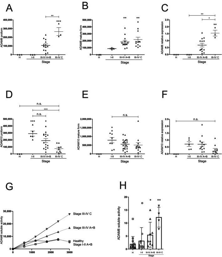
Aim: Periodontal disease is driven by oral pathogens, including Porphyromonas gingivalis, and the release of inflammatory cytokines. These cytokines (e.g., TNF) or their receptors (e.g., IL-1R) are substrates of a disintegrin and metalloproteinases (ADAMs). In this study, we aimed to determine the effects of ADAMs on periodontal disease phenotypes.

Materials and methods: Western blot and FRET-based activity measurements of the gingival crevicular fluid (GCF) of patients were compared with those of infected (P. gingivalis) or cytokine-stimulated oral keratinocytes and primary human neutrophils, respectively. This was accompanied by an analysis of the released extracellular vesicles and MMP9 activity.

Results: In the GCF of patients, ADAM8 protein expression and activity were correlated with disease stage, whereas ADAM10 protein expression was inversely correlated with disease stage. Infection and the resulting cytokine release orchestrated the release of soluble ADAM8 by oral keratinocytes and primary neutrophils as soluble ectodomain and on exosomes, respectively. Furthermore, ADAM8 regulated the release of ADAM10 and MMP9.

Conclusion: Dysregulation of cell-associated and extracellular ADAM proteolytic activity may be an essential regulatory element in the progression of periodontal disease driven by ADAM8. The influence of ADAM8 on disease onset and the evaluation of targeting ADAM8 as a potential and novel local treatment option should be addressed in future translational in vivo studies.

Keywords: cell–matrix interactions; infectious disease(s); matrix metalloproteinases; periodontal disease; proteases/proteinases.


Conflict of interest statement

The authors declare no conflicts of interest.

Figures

FIGURE 1
FIGURE 1
ADAM8 proteolytic activity in gingival fluid is associated with disease severity. (A–F) Sulcus fluid from patients with different disease stages were investigated by Western blotting to analyse the protein expression and maturation of ADAM8 and ADAM10 using antibodies against the C‐terminal domains. Band intensity was quantified by densitometry (A, pro‐form ADAM8; 120 kDa; B, mature form ADAM8, 90 kDa; D, pro‐form ADAM10; 100 kDa; E, mature form ADAM10, 70 kDa). Maturation was determined as ratio of the mature form and pro‐form of the respective protease (C and F). (G, H) Sulcus fluid was resuspended in activity buffer and subjected to ADAM8 activity measurement using a FRET‐based substrate (n = 3–12 per group). Activity over time is shown in (G) and was used for the calculation of the slope shown in (H). Data are shown as mean ± SD in (A–F) and (H), and activity over time in (G). Statistical analysis was performed with one‐way ANOVA followed by Tukey's post hoc test. Asterisks without line represent statistically significant differences to healthy patients; asterisks with line between groups (*p < 0.05, **p < 0.01, ***p < 0.001).
FIGURE 2
FIGURE 2
P. gingivalis induces ADAM8 expression and activity in human oral keratinocytes. Human primary keratinocytes (K2 cells) were grown until confluency and infected with P. gingivalis (2 h, 0.1 McF). (A–E) The cells were lysed, and the samples were analysed by Western blotting using antibodies against the C‐terminal domains of ADAM8 and ADAM10, respectively. GAPDH served as loading control. Band intensity was quantified by densitometry and normalized to uninfected cells. (F, G) The medium of the infected and non‐infected cells was subjected to differential centrifugation and density fractionation for exosome purification. The resulting pellets of different densities were analysed by Western blotting for the expression of ADAM8 and ADAM10 as well as Flottilin‐1 and CD9 as exosomal markers. Representative blots of three independent experiments are shown. (H, I) K2 cells were pre‐incubated with 10 μM BK‐1361, 10 μM GI254023X, or 0.01% DMSO (vehicle control) for 30 min and infected with P. gingivalis (2 h, 0.1 McF). The medium was analysed for ADAM8 or ADAM10 activity using a FRET‐based assay, and the slope of the activity over time was calculated. Quantitative data are shown as mean + SD of three independent experiments. Statistical analysis was performed with one‐sample t‐test (A–E) and one‐way ANOVA (H, I). Asterisks represent statistical differences (*p < 0.05, **p < 0.01, ***p < 0.001, ****p < 0.0001, n.s. not significant).
FIGURE 3
FIGURE 3
P. gingivalis‐induced ADAM8 activation correlates with proteolytic disbalance. (A, B) Primary human neutrophils were infected with P. gingivalis (2 h, 0.1 McF) or left uninfected. The medium of the infected and non‐infected cells was subjected to differential centrifugation and density fractionation for exosome purification. The resulting pellets of different density were analysed by Western blotting for the expression of ADAM8 and ADAM10 as well as Flottilin‐1 and CD9 as exosomal markers. Representative images of three independent experiments are shown. (C) Sulcus fluid from patients with different disease stages was analysed for MMP‐9 expression by ELISA. (D, E) Primary human neutrophils were pre‐incubated with 10 μM BK‐1361, 10 μM GI254023X or 0.01% DMSO (vehicle control) for 30 min and infected with P. gingivalis (2 h, 0.1 McF). Subsequently, the cells were lysed, and the cell lysate and the medium were analysed for MMP‐9 expression and release by ELISA. Neutrophil data are shown as mean ± SD of three independent experiments. Statistical analysis was performed using one‐way ANOVA followed by Tukey's post hoc test. Asterisks without line represent statistically significant differences with healthy patients and asterisks with line between groups (*p < 0.05, ***p < 0.001, ****p < 0.0001, n.s. not significant).
FIGURE 4
FIGURE 4
P. gingivalis‐induced ADAM8 activation correlates with tissue destruction. (A–D) K2 cells were grown until forming a monolayer, incubated with mitomycin (1 μg/mL) for 2 h followed by wound induction using an automatic scratcher. (A, B) Primary human neutrophils were infected with P. gingivalis (2 h, 0.1 McF). The medium of the infected cells was subjected to differential centrifugation and density fractionation for exosome purification and collection of soluble factors (supernatant). Subsequently, K2 cells were incubated with (A) exosomes or (B) soluble factors in the presence or absence of 10 μM BK‐1361 or 10 μM GI254023X and evaluated for wound closure. (C, D) Primary human neutrophils were pre‐incubated with 10 μM BK‐1361 or 0.01% DMSO (vehicle control) for 30 min and infected with P. gingivalis (2 h, 0.1 McF) followed by exosomes purification and collection of soluble factors from the medium. Subsequently, K2 cells were incubated with (C) exosomes or (D) soluble factors and evaluated for wound closure. Data are shown as a percentage of wound closure relative to closed wound. Quantitative data are shown as means ± SD of eight independent experiments. Asterisks indicate significance among treated cells calculated using one‐way ANOVA and Tukey's post‐test (*p < 0.05, **p < 0.01, ***p < 0.001).
FIGURE 5
FIGURE 5
Cytokines regulate ADAM8 expression in human oral keratinocytes. (A–E) K2 cells were grown until confluency and stimulated with a combination of TNFα, INF‐γ and IL‐1β (10 μM each) or sterile water (vehicle control) for 24 and 48 h. Subsequently, the cells were lysed, and the samples were analysed by Western blotting using antibodies against the C‐terminus of ADAM8 and ADAM10, respectively, with β‐actin as loading control. Band intensity was quantified by densitometry and normalized to unstimulated cells. Data are shown as mean + SD of three independent experiments. Statistical analysis was performed by one‐sample t‐test. Asterisks represent statistical differences (*p < 0.05, **p < 0.01, ***p < 0.001).
FIGURE 6
FIGURE 6
Regulation of extracellular release of ADAM8 and ADAM10 by cytokines. (A, B) K2 cells were grown until confluency and stimulated with a combination of TNFα, INF‐γ and IL‐1β (10 μM each) or sterile water (vehicle control) for 48 h. The medium of the stimulated and non‐stimulated cells was subjected to differential centrifugation and density fractionation for exosome purification. The resulting pellets of different density were analysed by Western blotting for the expression of ADAM8 and ADAM10 as well as Flottilin‐1 and CD9 as exosomal markers. Representative blots of three independent experiments are shown. (C–F) K2 cells (C, D) or primary human neutrophils (E, F) were pre‐incubated with 10 μM BK‐1361, 10 μM GI254023X or 0.01% DMSO (vehicle control) for 30 min followed by stimulation with a combination of TNFα, INF‐γ and IL‐1β (10 μM each) or sterile water (vehicle control) for 48 h. The medium was analysed for ADAM8 (C, E) or ADAM10 (D, F) activity using a FRET‐based activity assay. Data are shown as mean + SD of three independent experiments. Statistical analysis was performed using one‐sample t‐test. Asterisks without line represent statistically significant differences to healthy patients, and asterisks with line between groups (*p < 0.05, **p < 0.01, ***p < 0.001).

References

    1. Abdulkareem, A. A. , Al‐Taweel F. B., Al‐Sharqi A. J. B., Gul S. S., Sha A., and Chapple I. L. C.. 2023. “Current Concepts in the Pathogenesis of Periodontitis: From Symbiosis to Dysbiosis.” Journal of Oral Microbiology 15, no. 1: 2197779. 10.1080/20002297.2023.2197779. - DOI - PMC - PubMed
    1. Aljohmani, A. , Andres N. N., and Yildiz D.. 2022. “ Pseudomonas aeruginosa Alters Critical Lung Epithelial Cell Functions Through Activation of ADAM17.” Cells 11, no. 15: 2303. 10.3390/cells11152303. - DOI - PMC - PubMed
    1. Aljohmani, A. , Opitz B., Bischoff M., and Yildiz D.. 2022. “ Pseudomonas aeruginosa Triggered Exosomal Release of ADAM10 Mediates Proteolytic Cleavage in Trans.” International Journal of Molecular Sciences 23, no. 3: 1259. 10.3390/ijms23031259. - DOI - PMC - PubMed
    1. Aljohmani, A. , and Yildiz D.. 2020. “A Disintegrin and Metalloproteinase‐Control Elements in Infectious Diseases.” Frontiers in Cardiovascular Medicine 7: 608281. 10.3389/fcvm.2020.608281. - DOI - PMC - PubMed
    1. Aung, W. P. P. , Chotjumlong P., Pata S., et al. 2017. “Inducible Expression of a Disintegrin and Metalloproteinase 8 in Chronic Periodontitis and Gingival Epithelial Cells.” Journal of Periodontal Research 52, no. 3: 582–593. 10.1111/jre.12426. - DOI - PubMed

Publication types

MeSH terms

LinkOut - more resources