. 2024 Sep 24;10(3):e004600.
doi: 10.1136/rmdopen-2024-004600.

Genes deregulated in giant cell arteritis by Nanostring nCounter gene expression profiling in temporal artery biopsies

Affiliations

Genes deregulated in giant cell arteritis by Nanostring nCounter gene expression profiling in temporal artery biopsies

Ilaria Ferrigno et al. RMD Open. .

Abstract

Objective: To identify differentially expressed genes in temporal artery biopsies (TABs) from patients with giant cell arteritis (GCA) with different histological patterns of inflammation: transmural inflammation (TMI) and inflammation limited to adventitia (ILA), compared with normal TABs from patients without GCA.

Methods: Expression of 770 immune-related genes was profiled with the NanoString nCounter PanCancer Immune Profiling Panel on formalin-fixed paraffin-embedded TABs from 42 GCA patients with TMI, 7 GCA patients with ILA and 7 non-GCA controls.

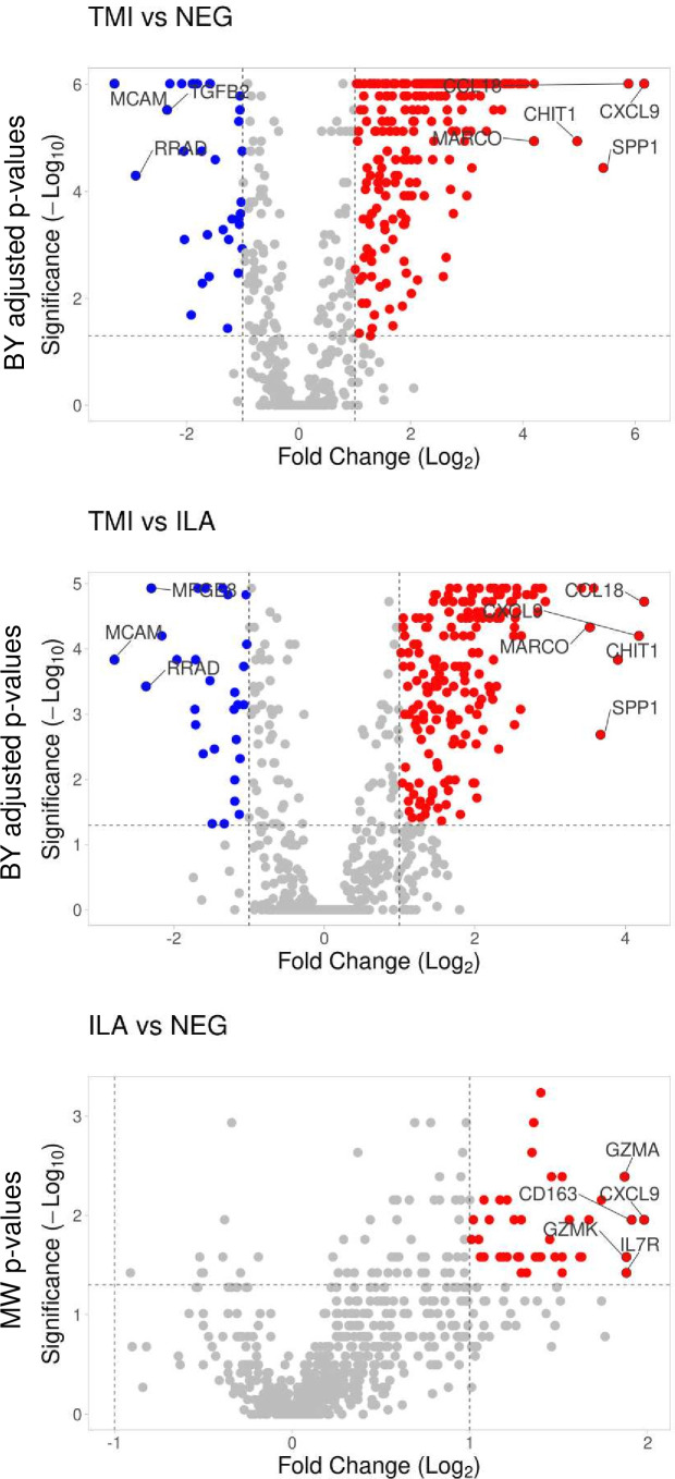
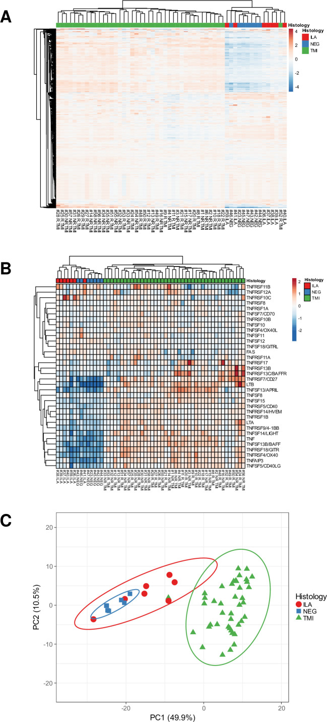
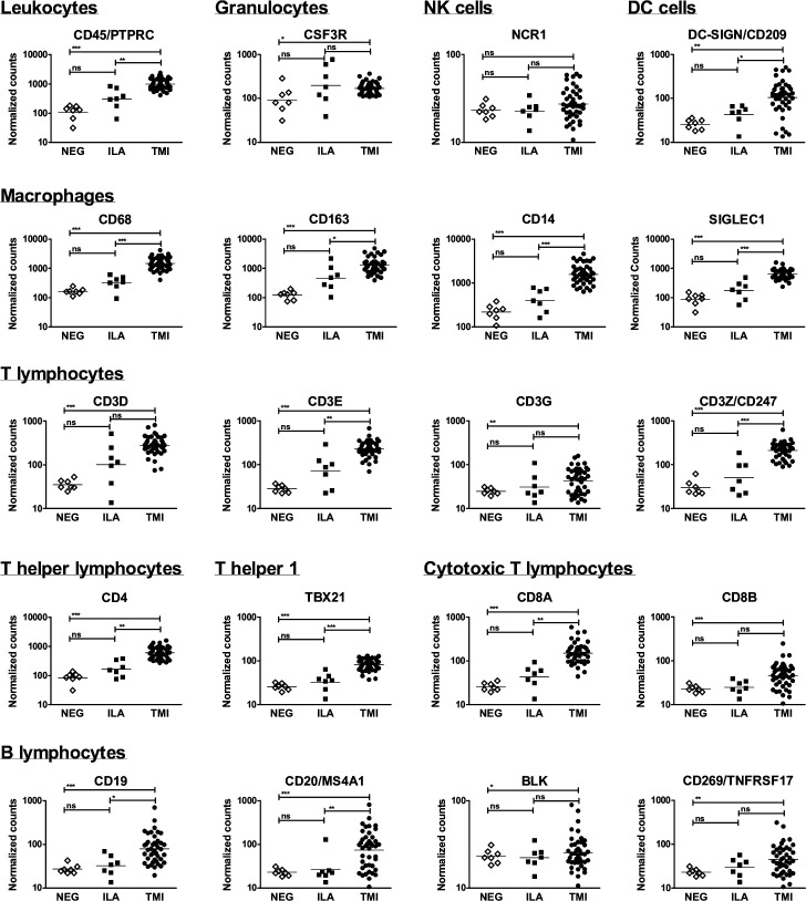
Results: Unsupervised clustering of the samples revealed two distinct groups: normal TABs and TABs with ILA in one group, 41/42 TABs with TMI in the other one. TABs with TMI showed 31 downregulated and 256 upregulated genes compared with normal TABs; they displayed 26 downregulated and 187 upregulated genes compared with TABs with ILA (>2.0 fold changes and adjusted p values <0.05). Gene expression in TABs with ILA resembled normal TABs although 38 genes exhibited >2.0 fold changes, but these changes lost statistical significance after Benjamini-Yekutieli correction. Genes encoding TNF superfamily members, immune checkpoints, chemokine and chemokine receptors, toll-like receptors, complement molecules, Fc receptors for IgG antibodies, signalling lymphocytic activation molecules, JAK3, STAT1 and STAT4 resulted upregulated in TMI.

Conclusions: TABs with TMI had a distinct transcriptome compared with normal TABs and TABs with ILA. The few genes potentially deregulated in ILA were also deregulated in TMI. Gene profiling allowed to deepen the knowledge of GCA pathogenesis.

Keywords: Autoimmune Diseases; Giant Cell Arteritis; Inflammation; Systemic vasculitis.


Conflict of interest statement

Competing interests: None declared.

Figures

Figure 1
Figure 1. Visualisation of the differentially expressed genes between the histological patterns of inflammation in TABs. Volcano plots show Log2 (fold changes of normalised gene counts) in the x-axis; −log10 (p values) in the y-axis. Red dots represent the upregulated genes with >2 fold changes and p values <0.05. Blue dots represent the downregulated genes with <−2 fold changes and p values <0.05. Benjamini-Yekutieli (BY) adjusted p values are shown for TMI versus NEG and TMI versus ILA. Uncorrected p values resulting from Mann-Whitney (MW) test are shown for ILA versus NEG. ILA, inflammation limited to adventitia; NEG, TABs without inflammation; TABs, temporal artery biopsies; TMI, transmural inflammation.
Figure 2
Figure 2. Unsupervised clustering of the samples. (A) Hierarchical clustering using the normalised expression of the 770 genes. (B) Hierarchical clustering using the subset of the TNF superfamily genes. (C) Principal component (PC) analysis using the normalised expression of the 770 genes. The x-axis and y-axis show principal component 1 and principal component 2. N=56 samples. ILA, inflammation limited to adventitia; NEG, TABs without inflammation; TMI, transmural inflammation.
Figure 3
Figure 3. Characterisation of the immune cell infiltrate in temporal artery biopsies. Normalised expression levels of transcripts reflecting T and B lymphocytes, macrophages, granulocytes, natural killer (NK) cells and dendritic cells (DCs), are shown in the group of samples. Lines indicate geometric means. Expression levels in the group of samples were compared with Kruskal-Wallis test with Dunn’s post-test, except for CD3G, CD8B, CD20, BLK, CD269 whose expressions were compared with Fisher’s exact test. *p<0.05; **p<0.01; ***p<0.001. ILA, inflammation limited to adventitia; NEG, TABs without inflammation; TMI, transmural inflammation.

References

    1. Salvarani C, Pipitone N, Versari A, et al. Clinical features of polymyalgia rheumatica and giant cell arteritis. Nat Rev Rheumatol. 2012;8:509–21. doi: 10.1038/nrrheum.2012.97. - DOI - PubMed
    1. Cavazza A, Muratore F, Boiardi L, et al. Inflamed temporal artery: histologic findings in 354 biopsies, with clinical correlations. Am J Surg Pathol. 2014;38:1360–70. doi: 10.1097/PAS.0000000000000244. - DOI - PubMed
    1. Régent A, Mouthon L. Treatment of Giant Cell Arteritis (GCA) J Clin Med. 2022;11:1799. doi: 10.3390/jcm11071799. - DOI - PMC - PubMed
    1. Weyand CM, Goronzy JJ. Immunology of Giant Cell Arteritis. Circ Res. 2023;132:238–50. doi: 10.1161/CIRCRESAHA.122.322128. - DOI - PMC - PubMed
    1. Samson M, Corbera-Bellalta M, Audia S, et al. Recent advances in our understanding of giant cell arteritis pathogenesis. Autoimmun Rev. 2017;16:833–44. doi: 10.1016/j.autrev.2017.05.014. - DOI - PubMed