Beyond visualizing the bird beak: esophagram, timed barium esophagram and manometry in achalasia and its 3 subtypes
- PMID: 39317828
- PMCID: PMC11947050
- DOI: 10.1007/s00261-024-04554-8
Beyond visualizing the bird beak: esophagram, timed barium esophagram and manometry in achalasia and its 3 subtypes
Abstract
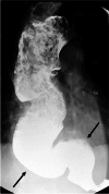
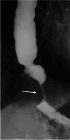
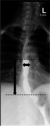
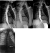
Achalasia is a rare esophageal motility disorder characterized by lack of primary peristalsis and a poorly relaxing lower esophageal sphincter. This disease process can be examined several ways and these evaluations can offer complementary information. There are three manometric subtypes of achalasia, with differing appearances on esophagram. Differentiating them is clinically important, because treatment for the subtypes varies. Timed barium esophagram (TBE) is a simple test to quantitatively evaluate esophageal emptying. TBE can be used to diagnose achalasia and assess treatment response. Considerable variation in the TBE protocol exist in the literature. We propose a standardized approach for TBE to allow for comparison across institutions.
Keywords: Achalasia; Esophagram; Manometry; Timed barium esophagram.
© 2024. The Author(s).
Conflict of interest statement
Declarations. Conflict of interest: The authors declare no competing interests.
Figures


Similar articles
-
Diagnostic Accuracy of Timed Barium Esophagram for Achalasia.Gastroenterology. 2025 Jul;169(1):63-72. doi: 10.1053/j.gastro.2025.02.013. Epub 2025 Feb 26. Gastroenterology. 2025. PMID: 40020937
-
Assessment of Esophageal Emptying in Patients with Dysphagia: Differences Between High-Resolution Impedance Manometry and Timed Barium Esophagram.Acta Gastroenterol Belg. 2025 Apr-Jun;88(2):89-95. doi: 10.51821/88.1.13772. Acta Gastroenterol Belg. 2025. PMID: 40504575
-
Timed barium esophagram in achalasia types.Dis Esophagus. 2015 May-Jun;28(4):336-44. doi: 10.1111/dote.12212. Epub 2014 Mar 20. Dis Esophagus. 2015. PMID: 24649871
-
The history and use of the timed barium esophagram in achalasia, esophagogastric junction outflow obstruction, and esophageal strictures.Neurogastroenterol Motil. 2025 Jan;37(1):e14928. doi: 10.1111/nmo.14928. Epub 2024 Sep 30. Neurogastroenterol Motil. 2025. PMID: 39345000 Review.
-
Quantitative methods to determine efficacy of treatment in achalasia.Gastrointest Endosc Clin N Am. 2001 Apr;11(2):409-24, viii-ix. Gastrointest Endosc Clin N Am. 2001. PMID: 11319070 Review.
Cited by
-
Esophageal motility disorders other than achalasia.Abdom Radiol (NY). 2025 Sep;50(9):3993-4003. doi: 10.1007/s00261-025-04828-9. Epub 2025 Mar 1. Abdom Radiol (NY). 2025. PMID: 40024921 Review.
References
-
- Momodu, II and J.M. Wallen, Achalasia, in StatPearls. 2024: Treasure Island (FL).
Publication types
MeSH terms
Substances
LinkOut - more resources
Full Text Sources

