. 2024 Sep;28(18):e70114.
doi: 10.1111/jcmm.70114.

Inhibition of HDAC8 mitigates AKI by reducing DNA damage and promoting homologous recombination repair

Affiliations

Inhibition of HDAC8 mitigates AKI by reducing DNA damage and promoting homologous recombination repair

Yanjin Wang et al. J Cell Mol Med. 2024 Sep.

Abstract

Nephrotoxicity is a major side effect of platinum-based antineoplastic drugs, and there is currently no available therapeutic intervention. Our study suggests that targeting histone deacetylase 8 could be a potential treatment for cisplatin-induced acute kidney injury (AKI). In a murine model of AKI induced by cisplatin, the administration of PCI-34051, a selective inhibitor of HDAC8, resulted in significant improvement in renal function and reduction in renal tubular damage and apoptosis. Pharmacological inhibition of HDAC8 also decreased caspase-3 and PARP1 cleavage, attenuated Bax expression and preserved Bcl-2 levels in the injured kidney. In cultured murine renal epithelial cells (mRTECs) exposed to cisplatin, treatment with PCI-34051 or transfection with HDAC8 siRNA reduced apoptotic cell numbers and diminished expression of cleaved caspase-3 and PARP1; conversely, overexpression of HDAC8 intensified these changes. Additionally, PCI-34051 reduced p53 expression levels along with those for p21, p-CDK2 and γ-H2AX while preserving MRE11 expression in the injured kidney. Similarly, pharmacological and genetic inhibition of HDAC8 reduced γ-H2AX and enhanced MRE11 expression; conversely, HDAC8 overexpression exacerbated these changes in mRTECs exposed to cisplatin. These results support that HDAC8 inhibition attenuates cisplatin-induced AKI through a mechanism associated with reducing DNA damage and promoting its repair.

Keywords: DNA damage; HDAC8; acute kidney injury; apoptosis; cisplatin; homologous recombination repair.


Conflict of interest statement

The authors declare no conflicts of interest.

Figures
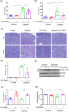
FIGURE 1
FIGURE 1
Inhibition of HDAC8 attenuates cisplatin‐induced AKI (A) Blood urea nitrogen (BUN) levels were measured using a colorimetric method in each group of mice. (B) Serum creatinine levels were detected by colorimetric method in each group of mice. (C) Periodic acid‐Schiff (PAS) staining revealed kidney structure damage in mice from each group. Scale bar = 100 μm. (D) Tubule injury scores of renal tissue were assessed in each group. (E) Western blot analysis was performed to detect the expression of lysine acetylated proteins and HDAC8 in kidney tissues from mice in each group. (F) The relative abundance of lysine acetylated proteins in immunoblots was quantified by densitometric ratios of lysine acetylated proteins/Tubulin. (G) The relative abundance of HDAC8 in immunoblots was quantified by densitometric ratios of HDAC8/Tubulin. Data are presented as mean ± standard deviations. *p < 0.05, **p < 0.01.
FIGURE 2
FIGURE 2
HDAC8 inhibition reduces NGAL expression levels. (A) Western blot analysis was performed to detect the expression of NGAL in kidney tissues of mice from each group. (B) Relative abundance of NGAL protein expression levels compared to Tubulin. (C) Immunofluorescence staining revealed the expression of NGAL in kidney tissue from each group. Scale bar = 100 μm. (D) Comparison of the number of cells positive for NGAL immunofluorescence staining in each group. Data are presented as mean ± standard deviations. *p < 0.05, **p < 0.01.
FIGURE 3
FIGURE 3
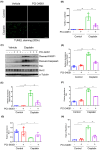
Inhibition of HDAC8 attenuates apoptosis of renal tubular epithelial cells. (A) TUNEL staining was performed on kidney tissue sections from mice in each group. Scale bar = 100 μm. (B) Statistical analysis showing the number of TUNEL‐staining positive cells in each group. (C) Western blot analysis was used to detect the expression of cleaved PARP1, cleaved caspase‐3, Bax and Bcl‐2 in the kidney tissues from mice in each group. (D–G) Relative abundance of cleaved PARP1, cleaved caspase‐3, Bax and Bcl‐2 protein expression levels compared to Tubulin. Data are expressed as mean ± standard deviations.*p < 0.05, **p < 0.01.
FIGURE 4
FIGURE 4
HDAC8 blockade alleviates renal tubular epithelial cell apoptosis and increases cell viability. (A) TUNEL staining of renal tubular epithelial cells in each group. Scale bar = 100 μm. (B) Comparison of the number of TUNEL‐staining positive cells in each group. (C) CCK8 was used to detect cell viability in each group. (D) Western blotting to detect the expression of cleaved PARP1, cleaved caspase‐3, and lysine acetylated histone of renal tubular epithelial cell in each group. (E) Relative abundance of cleaved PARP1 protein expression levels. (F) Relative abundance of cleaved caspase‐3 protein expression levels. (G) Relative abundance of lysine acetylated histone protein. Data are expressed as mean ± standard deviations. *p < 0.05, **p < 0.01, ***p < 0.001.
FIGURE 5
FIGURE 5
HDAC8 exacerbates histone deacetylation and promotes cisplatin‐induced apoptosis in renal tubular epithelial cells. (A) The protein levels of cleaved PARP1, cleaved caspase‐3, and acetylated histone protein in renal tubular epithelial cells in each group. Cells were treated with HDAC8 siRNA and control siRNA for 48 h, and then cultured in cisplatin‐containing medium for 24 h. (B) Relative abundance of cleaved PARP1 protein expression levels. (C) Relative abundance of cleaved caspase‐3 protein expression levels. (D) Relative abundance of lysine acetylated histone protein. (E) CCK8 was used to detect cell viability in each group. (F) Western blotting to detect the expression changes of the corresponding proteins of HDAC8 overexpression and control renal tubular epithelial cells in each group. (G) Relative abundance of cleaved PARP1 protein expression levels. (H) Relative abundance of cleaved caspase‐3 protein expression levels. (I) Relative abundance of lysine acetylated histone. (J): CCK8 was used to detect cell viability in each group. Data are expressed as mean ± standard deviations. *p < 0.05, **p < 0.01, ***p < 0.001.
FIGURE 6
FIGURE 6
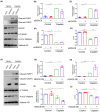
HDAC8 inhibition induces changes in the p53 signalling pathway. (A) The protein levels of p53, p21, p‐CDK2 and CDK2 in kidney tissue from each group are shown. (B) Relative abundance of p53 protein expression levels. (C) Relative abundance of p21 protein expression levels. (D) Relative abundance of p‐CDK2 protein expression levels. Data are presented as mean ± standard deviations. *p < 0.05, **p < 0.01.
FIGURE 7
FIGURE 7
HDAC8 inhibition enhances homologous recombination repair. (A) Western blot analysis of γ‐H2AX protein levels in kidney tissues from each group. (B) Statistical analysis of relative abundance of γ‐H2AX expression levels. (C) Western blot analysis of MRE11 protein levels in kidney tissues from each group. (D) Statistical analysis of relative abundance of MRE11 expression levels. (E) Western blot analysis of γ‐H2AX and MRE11 protein levels in renal tubular epithelial cells from each group. (F) Statistical analysis of relative abundance of γ‐H2AX expression levels. (G) Statistical analysis of relative abundance of MRE11 expression levels. (H) Protein levels of γ‐H2AX and MRE11 in HDAC8 overexpressing renal tubular epithelial cells from each group. (I) Statistical analysis of relative abundance of γ‐H2AX expression levels. (J) Statistical analysis on the relative abundanceof MRE11expressionlevels. Data are presented as mean ± standard deviation. *p < 0.05, **p < 0.01.

Similar articles

References

    1. Tang C, Livingston MJ, Safirstein R, Dong Z. Cisplatin nephrotoxicity: new insights and therapeutic implications. Nat Rev Nephrol. 2023;19(1):53‐72. doi:10.1038/s41581-022-00631-7 - DOI - PubMed
    1. Wang J, Shen F, Liu F, Zhuang S. Histone modifications in acute kidney injury. Kidney Dis (Basel). 2022;8(6):466‐477. doi:10.1159/000527799 - DOI - PMC - PubMed
    1. Zhou X, Chen H, Shi Y, Ma X, Zhuang S, Liu N. The role and mechanism of histone deacetylases in acute kidney injury. Front Pharmacol. 2021;12:695237. doi:10.3389/fphar.2021.695237 - DOI - PMC - PubMed
    1. Tang J, Zhuang S. Epigenetics in acute kidney injury. Curr Opin Nephrol Hypertens. 2015;24(4):351‐358. doi:10.1097/MNH.0000000000000140 - DOI - PMC - PubMed
    1. Ellmeier W, Seiser C. Histone deacetylase function in CD4(+) T cells. Nat Rev Immunol. 2018;18(10):617‐634. doi:10.1038/s41577-018-0037-z - DOI - PubMed

MeSH terms

LinkOut - more resources