. 2024 Nov 18;45(6):1175-1187.
doi: 10.24272/j.issn.2095-8137.2024.219.

IDH2 and GLUD1 depletion arrests embryonic development through an H4K20me3 epigenetic barrier in porcine parthenogenetic embryos

Affiliations

IDH2 and GLUD1 depletion arrests embryonic development through an H4K20me3 epigenetic barrier in porcine parthenogenetic embryos

Cheng-Lin Zhan et al. Zool Res. .

Abstract

Isocitrate dehydrogenase 2 (IDH2) and glutamate dehydrogenase 1 (GLUD1) are key enzymes involved in the production of α-ketoglutarate (α-KG), a metabolite central to the tricarboxylic acid cycle and glutamine metabolism. In this study, we investigated the impact of IDH2 and GLUD1 on early porcine embryonic development following IDH2 and GLUD1 knockdown (KD) via double-stranded RNA (dsRNA) microinjection. Results showed that KD reduced α-KG levels, leading to delayed embryonic development, decreased blastocyst formation, increased apoptosis, reduced blastomere proliferation, and pluripotency. Additionally, IDH2 and GLUD1 KD induced abnormally high levels of trimethylation of lysine 20 of histone H4 (H4K20me3) at the 4-cell stage, likely resulting in transcriptional repression of embryonic genome activation (EGA)-related genes. Notably, KD of lysine methyltransferase 5C ( KMT5C) and supplementation with exogenous α-KG reduced H4K20me3 expression and partially rescued these defects, suggesting a critical role of IDH2 and GLUD1 in the epigenetic regulation and proper development of porcine embryos. Overall, this study highlights the significance of IDH2 and GLUD1 in maintaining normal embryonic development through their influence on α-KG production and subsequent epigenetic modifications.

异柠檬酸脱氢酶2(IDH2)和谷氨酸脱氢酶1(GLUD1)是参与α-酮戊二酸(α-KG)生成的关键酶,α-KG是三羧酸循环和谷氨酰胺代谢中的重要代谢物。在该研究中,我们通过显微注射dsRNA敲低了IDH2和GLUD1的表达,重点研究了它们对早期猪胚胎发育的影响。敲低导致4细胞及囊胚期猪胚胎胞内α-KG水平降低,进而引起胚胎发育延迟、囊胚形成率下降、细胞凋亡增加、卵裂球增殖和多能性降低。此外,IDH2和GLUD1的敲低导致在4细胞阶段组蛋白H4赖氨酸20三甲基化(H4K20me3)水平异常升高,可能导致与胚胎基因组激活(EGA)相关基因的转录抑制。重要的是,赖氨酸甲基转移酶5C(KMT5C)的敲低和外源性α-KG的补充降低了H4K20me3的表达,部分恢复了这些缺陷,表明IDH2和GLUD1在表观遗传调控和猪胚胎正常发育中的关键作用。该研究强调了IDH2和GLUD1在通过影响α-KG的生成及其后续表观遗传修饰来维持正常胚胎发育中的重要性。.

Keywords: A-ketoglutarate; Embryonic development; GLUD1; H4K20me3; IDH2.


Conflict of interest statement

The authors declare that they have no competing interests.

Figures

Figure 1
Figure 1
IDH2 and GLUD1 mRNA and protein expression levels during early porcine embryonic development A: Relative expression of IDH2 mRNA in 2-cell (2C), 4-cell (4C), morula (MO), and blastocyst (BL) stage embryos. B: Representative immunofluorescence (IF) images of IDH2 protein from 2C to BL stages. C: Relative fluorescence intensity of IDH2 protein. D: Relative expression of GLUD1 mRNA from 2C to BL stages. E: Representative IF images of GLUD1 protein from 2C to BL stages. F: Relative fluorescence intensity of GLUD1 protein. Scale bar: 20 μm. ns: Not significant; *: P<0.05; **: P<0.01; ***: P<0.001; ****: P<0.0001.
Figure 2
Figure 2
Effects of IDH2 and GLUD1 knockdown (KD) on intracellular α-ketoglutarate (α-KG) content A: Relative expression of GLUD1 mRNA in 4C stage embryos following injection of IDH2 and GLUD1 dsRNA mix (DKD). Nuclear-free water was injected into porcine parthenotes as the control group (NC). B: Representative immunofluorescence (IF) staining images of GLUD1 protein. C: Relative fluorescence intensity of GLUD1 protein. D: Relative expression of IDH2 mRNA in NC and DKD 4C stage embryos. E: Representative IF images of IDH2 protein and Mito-Tracker Red CMXRos. F: Relative fluorescence intensity of IDH2 protein and Mito-Tracker Red CMXRos. G: Representative western blot images of IDH2 and GLUD1; GAPDH was used as the internal reference protein. H: Relative protein levels of IDH2 and GLUD1 in NC and DKD 4C stage embryos. I: Changes in intracellular α-ketoglutarate (α-KG) content in NC and DKD 4C and BL stage embryos. Scale bar: 20 μm. ns: Not significant; *: P<0.05; **: P<0.01; ***: P<0.001; ****: P<0.0001.
Figure 3
Figure 3
Effects of IDH2 and GLUD1 knockdown (KD) on porcine EGA A: Proportion of 1-cell (1C), 2C, 4C, 8-cell (8C), MO, and BL stage embryos to all embryos at 0, 24, 48, 72, 96, 120, and 144 h post-KD. B: Relative mRNA expression of embryonic genome activation (EGA)-related genes ( DPPA2, WEE1, EIF1A, RIF1, and ZSCAN4) in NC and DKD 4C stage embryos. C: Representative IF images of early transcription product SIRT1 in NC and DKD 4C stage embryos. Scale bars: 20 μm. D: Changes in fluorescence intensity at yellow line of SIRT1 protein and DAPI. ns: Not significant; *: P<0.05; **: P<0.01; ***: P<0.001; ****: P<0.0001.
Figure 4
Figure 4
Effects of IDH2 and GLUD1 knockdown (KD) on porcine BL quality A: BL rate in NC and DKD groups. B: Representative IF images of γH2Ax in NC and DKD BLs. C: Ratio of γH2Ax positive/total nuclei in NC and DKD BLs. D: Representative IF images of caspase-3 (CASP3) protein and EdU staining in NC and DKD BLs. E: Relative fluorescence intensity of CASP3 protein in NC and DKD BLs. F: Ratio of EdU positive/total nuclei in NC and DKD BLs. G: Representative IF images of OCT4 protein in NC and DKD BLs. H: Ratio of OCT4 positive/total nuclei in NC and DKD BLs. I: Total blastomere number in NC and DKD groups. J: BL diameter in NC and DKD groups. Scale bar: 20 μm. ns: Not significant; *: P<0.05; **: P<0.01; ***: P<0.001; ****: P<0.0001.
Figure 5
Figure 5
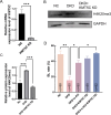
Effects of IDH2 and GLUD1 knockdown (KD) and dimethyl-α-KG (DM-α-KG) supplementation on histone modifications in porcine embryos A: Embryonic developmental rates in NC, DKD, DKD+1 μmol/L DM-α-KG, DKD+5 μmol/L DM-α-KG, DKD+10 μmol/L DM-α-KG, and DKD+20 μmol/L DM-α-KG groups. B: Representative western blot images in 4C stage embryos of NC, DKD, and DKD+10 μmol/L DM-α-KG (DKD+α-KG) groups; GAPDH was used as the internal reference protein. C: Relative protein levels in 4C stage embryos of NC, DKD, and DKD+α-KG groups. D: Representative IF images of H4K20me3 in 4C stage embryos of NC, DKD, and DKD+α-KG groups. Scale bar: 20 μm. E: Fluorescence intensity of H4K20me3 at yellow line in 4C stage embryos of NC, DKD, DKD+α-KG groups. ns: Not significant; *: P<0.05; **: P<0.01; ***: P<0.001; ****: P<0.0001.
Figure 6
Figure 6
Effects of IDH2 and GLUD1 knockdown (KD) on H4K20me3 levels in porcine embryos A: α-KG is a cofactor of H4K20me3 demethylase RAD23. B: Representative western blot images of H4K20me3 in 1C, 2C, 4C, and BL stage embryos; GAPDH was used as the internal reference protein. C: Relative H4K20me3 protein levels in 1C, 2C, 4C, and BL stage embryos. D: Representative IF images of H4K20me3 in NC and DKD 1C, 2C, 4C, MO, and BL stage embryos. Scale bars: 20 μm. E: Relative fluorescence intensity of H4K20me3 in NC and DKD 1C, 2C, 4C, MO, and BL stage embryos. ns: Not significant; *: P<0.05; **: P<0.01; ***: P<0.001; ****: P<0.0001.
Figure 7
Figure 7
Effects of KMT5C knockdown (KD) and DM-α-KG supplementation on porcine embryonic development ability A: Relative expression of KMT5C mRNA in NC and KMT5C KD embryos. B: Representative western blot images of H4K20me3 protein in NC, DKD, and DKD+KMT5C KD embryos at 4C stage. C: Relative H4K20me3 protein levels in NC, DKD, and DKD+KMT5C KD embryos at 4C stage. D: BL formation rate in NC, DKD, DKD+α-KG, DKD+KMT5C KD, and DKD+α-KG+KMT5C KD groups. ns: Not significant; *: P<0.05; **: P<0.01; ***: P<0.001; ****: P<0.0001.
Figure 8
Figure 8
Effects of KMT5C knockdown (KD) and DM-α-KG supplementation on porcine blastocyst (BL) quality A: Representative IF images of γH2Ax in NC, DKD, DKD+α-KG, DKD+KMT5C KD, and DKD+α-KG+KMT5C KD BLs. B: Ratio of γH2Ax positive/total nuclei in NC, DKD, DKD+α-KG, DKD+KMT5C KD, and DKD+α-KG+KMT5C KD BLs. C: Representative IF images of CASP3 protein and EdU staining in NC, DKD, DKD+α-KG, DKD+KMT5C KD, and DKD+α-KG+KMT5C KD BLs. D: Relative fluorescence intensity of CASP3 protein in NC, DKD, DKD+α-KG, DKD+KMT5C KD, and DKD+α-KG+KMT5C KD BLs. CASP3: Caspase-3. E: Ratio of EdU positive/total nuclei in NC, DKD, DKD+α-KG, DKD+KMT5C KD, and DKD+α-KG+KMT5C KD BLs. F: Representative IF images of OCT4 protein in NC, DKD, DKD+α-KG, DKD+KMT5C KD, and DKD+α-KG+KMT5C KD BLs. G: Ratio of OCT4 positive/total nuclei in NC, DKD, DKD+α-KG, DKD+KMT5C KD, and DKD+α-KG+KMT5C KD BLs. Scale bar: 20 μm. ns: Not significant; *: P<0.05; **: P<0.01; ***: P<0.001; ****: P<0.0001.
Figure 9
Figure 9
Schematic of effects of IDH2 and GLUD1 knockdown (KD) on porcine embryonic development The IDH2 and GLUD1 enzymes produce intracellular α-KG. Hence, IDH2 and GLUD1 KD results in depletion of intracellular α-KG content, which impairs H4K20me3 demethylation. Thus, intracellular α-KG depletion impairs embryonic development through transcriptional repression as a result of high H4K20me3 levels.

References

    1. Adamkova K, Yi YJ, Petr J, et al SIRT1-dependent modulation of methylation and acetylation of histone H3 on lysine 9 (H3K9) in the zygotic pronuclei improves porcine embryo development. Journal of Animal Science and Biotechnology. 2017;8:83. doi: 10.1186/s40104-017-0214-0. - DOI - PMC - PubMed
    1. Al-Khallaf H Isocitrate dehydrogenases in physiology and cancer: biochemical and molecular insight. Cell & Bioscience. 2017;7:37. - PMC - PubMed
    1. Antony J, Oback F, Chamley LW, et al Transient JMJD2B-mediated reduction of H3K9me3 levels improves reprogramming of embryonic stem cells into cloned embryos. Molecular and Cellular Biology. 2013;33(5):974–983. doi: 10.1128/MCB.01014-12. - DOI - PMC - PubMed
    1. Balakrishnan L, Milavetz B Decoding the histone H4 lysine 20 methylation mark. Critical Reviews in Biochemistry and Molecular Biology. 2010;45(5):440–452. doi: 10.3109/10409238.2010.504700. - DOI - PubMed
    1. Cao XW, Chen YR, Wu B, et al. 2020. Histone H4K20 demethylation by two hHR23 proteins. Cell Reports, 30(12): 4152–4164. e6.

MeSH terms

LinkOut - more resources