Review
. 2024 Mar 23;34(4):688-701.
doi: 10.1055/s-0044-1781459. eCollection 2024 Oct.

Anal and Perianal Masses: The Common, the Uncommon, and the Rare

Affiliations
Review

Anal and Perianal Masses: The Common, the Uncommon, and the Rare

Antony Augustine et al. Indian J Radiol Imaging. .

Abstract

A variety of tumors involve the anal canal because the anal canal forms the transition between the digestive system and the skin, and this anatomical region is made of a variety of different cells and tissues. Magnetic resonance imaging (MRI) is the modality of choice for diagnosis and local staging of the anal canal and perianal neoplasms. In this pictorial review, we demonstrate the MRI anatomy of the anal canal and perianal region and display the imaging spectrum of tumors in the region along with an overview of its management. Imaging appearances of many tumorlike lesions that can cause diagnostic dilemmas are also demonstrated with pointers to differentiate between them.

Keywords: MRI; anal; cancer; neoplasms; perianal.


Conflict of interest statement

Conflict of Interest None declared.

Figures

Fig. 1
Fig. 1
( A ) Sagittal image of the anal canal demonstrating the planes to obtain axial ( yellow dashed line ) and coronal ( red dashed line ) T2 high-resolution images of the anal canal. ( B,C ) Magnetic resonance imaging (MRI) of the anatomy of the anal canal in coronal plane.
Fig. 2
Fig. 2
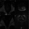
Anal canal squamous cell carcinoma (SCC) before treatment ( A,B ) and posttreatment ( C,D ). ( A ) Axial T2-weighted image (T2WI) and ( B ) high b -value diffusion weighted imaging (DWI) show typical appearance of treatment naïve anal canal SCC, which is Infiltrative, usually a circumferentially spreading lesion, which is intermediate on T2 and shows restricted diffusion. Postchemoradiotherapy (CRT) T2WIs in ( C ) axial and ( D ) high b-value DWI show complete response to CRT indicated by loss of bulk of lesion with T2 hypointensity signal without restricted diffusion.
Fig. 3
Fig. 3
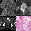
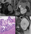
Mucinous adenocarcinoma of anal canal. ( A ) T2-weighted image (T2WI) and ( B ) short tau inversion recovery (STIR) image in axial plane show T2/STIR markedly hyperintense lobulated mass in the anal canal. ( C ) Axial computed tomography (CT) image through the anal canal done for planning radiotherapy (RT) shows the low attenuation of mucin. ( D ) Hematoxylin and eosin–stained histology microphotograph, of original magnification 40x, shows mucin lakes within the specimen.
Fig. 4
Fig. 4
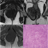
Anal melanoma. T2-weighted image (T2WI) in ( A ) axial and ( B ) coronal planes and ( C ) T1WI in axial plane show a T2 intermediate and T1 hyperintense lesion in the anal canal infiltrating the sphincter. ( D ) Hematoxylin and eosin–stained histology microphotograph, of original magnification 40x, shows melanin containing tumor cells.
Fig. 5
Fig. 5
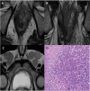
A human immunodeficiency virus (HIV) positive man with anal lymphoma. T2-weighted image (T2WI) in ( A ) axial and ( B ) coronal planes through the anal canal show an infiltrative T2 mildly hyperintense mass in the anal canal. ( C ) T2WI in axial plane through the inguinal level shows bilateral disproportionately large homogenous inguinal nodes. ( D ) Hematoxylin and eosin–stained histology microphotograph, of original magnification 100x, shows a tumor with plasmablastic cells suggestive of plasmablastic lymphoma.
Fig. 6
Fig. 6
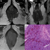
Anal leiomyoma. T2-weighted image (T2WI) in ( A ) axial and ( B ) coronal planes and ( C ) T1WI in axial plane show an exophytic growing smooth marginated T2 hypointense and T1 isointense lesion extending into the right ischioanal fossa. ( D ) Hematoxylin and eosin–stained histology microphotograph, of original magnification 40x, shows well-differentiated smooth muscle cells with red fibrillar cytoplasm showing no nuclear atypia.
Fig. 7
Fig. 7
Anal leiomyosarcoma. ( A ) T2-weighted image (T2WI), ( B ) short tau inversion recovery (STIR), and ( C ) T1-wegihted image (T1WI) in axial planes show as lobulated exophytic growing lesion with irregular margins with T2 heterogeneous signal due to necrosis. ( D ) Hematoxylin and eosin–stained histology microphotograph, of original magnification 40x, shows smooth muscle cells with a prominent nuclear atypia ( arrow ).
Fig. 8
Fig. 8
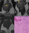
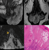
Anal gastrointestinal stromal tumor (GIST). T2-weighted image (T2WI) in ( A ) axial and ( B ) coronal planes and ( C ) T1WI in axial plane through the anal canal show exophytic growing smoothly marginated T1 isointense and T2 heterogeneous mass from the anal canal. The heterogeneity on T2 is due to multiple necrotic foci within the tumor ( yellow arrowheads in A and B ). ( D ) Hematoxylin and eosin–stained histology microphotograph, of original magnification 40x, shows bland spindle cells with elongated nuclei typical of spindle type of GIST.
Fig. 9
Fig. 9
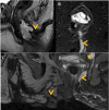
Malignancy associated with fistula-in-ano. T2-weighted image (T2WI) in ( A ) sagittal plane and ( B ) short tau inversion recovery (STIR) axial image through the anal canal show a T2/STIR markedly hyperintense lobulated mass in the anal canal with a high trans-sphincteric fistula-in-ano ( arrowheads in A and B ). The presence of a lesion at the internal opening of the fistula favors this to be a case of a fistula developing due to anal malignancy (malignancy presenting as a fistula-in-ano). Companion images of a case of malignancy developing in chronic fistula, which is seen on ( C ) T2 image and ( D ) STIR image as a complex trans-sphincteric fistula with multifocal areas of nodular soft tissue within its wall ( yellow arrowheads in C and D ). A heterogeneous enlarged mesorectal node is also seen ( yellow star in C ).
Fig. 10
Fig. 10
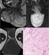
Aggressive angiomyxoma. T2-weighted image (T2WI) in ( A ) sagittal plane, ( B ) short tau inversion recovery (STIR) image in coronal plane, and ( C ) T1 image in axial plane show T2/STIR markedly hyperintense lobulated mass in the anal and perianal region with a characteristic laminated appearance. The lesion is hypointense on T1WI. ( D ) Hematoxylin and eosin–stained histology microphotograph, of original magnification 40x, shows myxoid stroma with scattered stellate to spindle cells.
Fig. 11
Fig. 11
Developmental cysts. ( A ) Tailgut cysts are multiloculated cysts in the retro-anal location, best demonstrated on T2 image. ( B ) The high protein content within the cyst can be seen as T1 fat-suppressed sequence (T1FS) hyperintensity. ( C ) Hematoxylin and eosin–stained histology microphotograph, of original magnification 40x, showing tailgut cyst lined by a columnar epithelium ( arrowhead ) and squamous epithelium ( arrow ). ( D ) Epidermoid cysts are unilocular on T2 images with the presence of T2 hypointense spheres within it representing keratin debris.
Fig. 12
Fig. 12
Perianal squamous cell carcinoma (SCC). T2- weighted image (T2WI) in ( A ) sagittal, ( B ) axial, and ( C ) coronal planes show T2 intermediate signal thickening in the posterior perianal skin, which reaches up to the anal verge ( arrowhead in A ). The diagnosis was done by biopsy and magnetic resonance imaging (MRI) was done as part of staging workup to evaluate anal canal involvement and to look for regional nodes. ( D ) Hematoxylin and eosin–stained histology microphotograph, of original magnification 40x, shows marked keratinization and minimal nuclear pleomorphism in a well-differentiated SCC.
Fig. 13
Fig. 13
Verrucous carcinoma at perianal region. T2-weighted image (T2WI) in ( A ) sagittal and ( B ) coronal planes and ( C ) short tau inversion recovery (STIR) image in axial plane shows a cauliflower like T2/STIR hyperintense mass in the perianal region with infiltration of the superficial and subcutaneous bundles of external anal sphincter ( arrowhead ). ( D ) Hematoxylin and eosin–stained histology microphotograph, of original magnification 40x, shows massive hyperkeratosis and parakeratosis.
Fig. 14
Fig. 14
Tumorlike lesions.
Fig. 15
Fig. 15
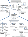
Diagnostic algorithm for an imaging-based diagnosis of lesions in and around the anal canal.

Similar articles

References

    1. Siegel R L, Miller K D, Fuchs H E, Jemal A. Cancer statistics, 2022. CA Cancer J Clin. 2022;72(01):7–33. - PubMed
    1. Solan P, Davis B. Anorectal anatomy and imaging techniques. Gastroenterol Clin North Am. 2013;42(04):701–712. - PubMed
    1. Brown G, Daniels I R, Richardson C, Revell P, Peppercorn D, Bourne M. Techniques and trouble-shooting in high spatial resolution thin slice MRI for rectal cancer. Br J Radiol. 2005;78(927):245–251. - PubMed
    1. Chandramohan A et al.MRI staging of anorectal malignancy—a reporting dilemma: is it adenocarcinoma or squamous cell carcinoma? J Gastrointestinal Abdominal Radiol. 2023;6:136–145.
    1. deSouza N M, Williams A D, Gilderdale D J. High-resolution magnetic resonance imaging of the anal sphincter using a dedicated endoanal receiver coil. Eur Radiol. 1999;9(03):436–443. - PubMed

LinkOut - more resources