

Ultrasound-Based Noncontrast Microvascular Imaging for Evaluation of Breast Lesions: Imaging Techniques and Review of Diagnostic Criteria
- PMID: 39318571
- PMCID: PMC11419773
- DOI: 10.1055/s-0044-1782162
Ultrasound-Based Noncontrast Microvascular Imaging for Evaluation of Breast Lesions: Imaging Techniques and Review of Diagnostic Criteria
Abstract
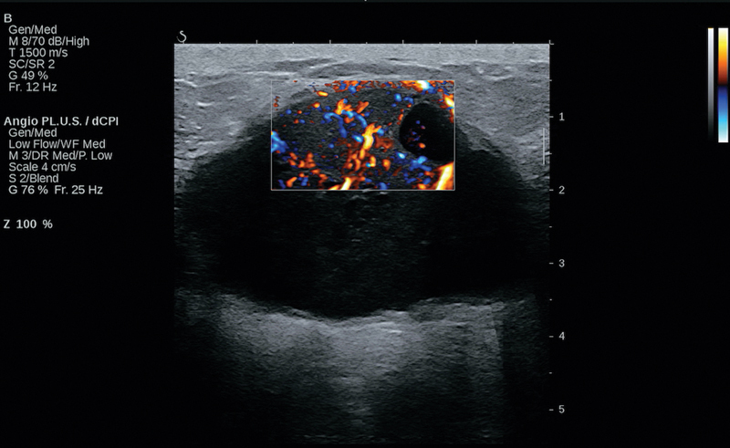
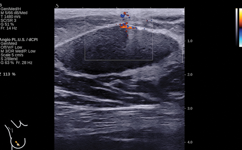
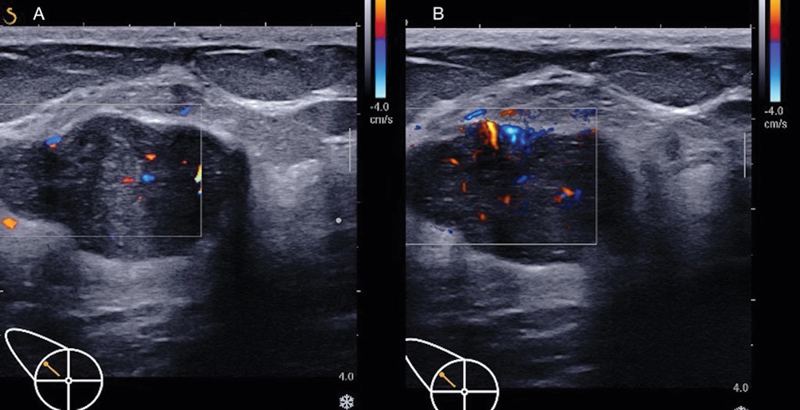
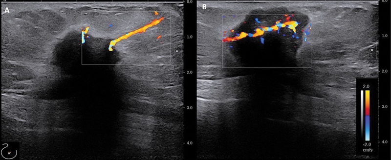
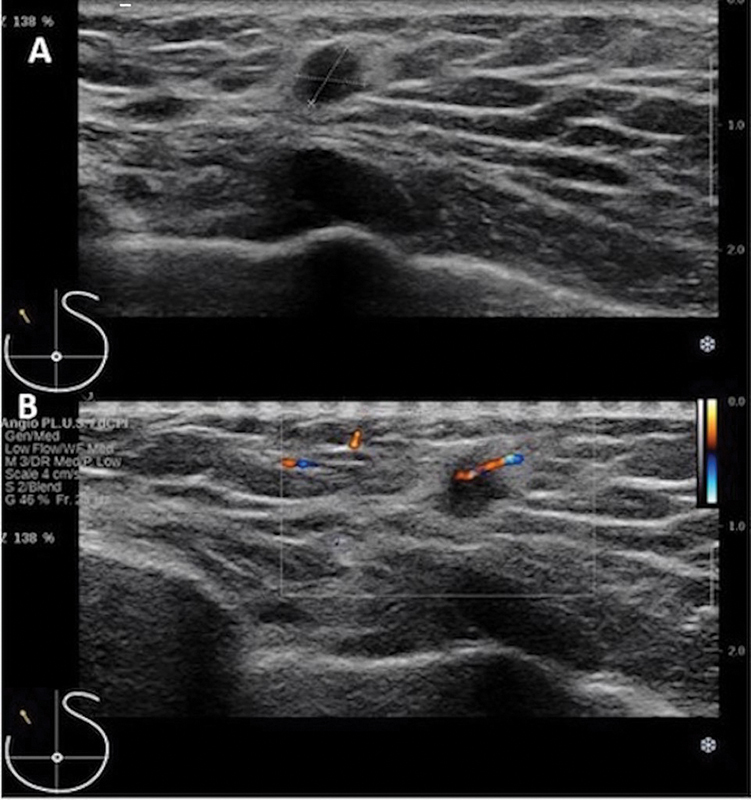
Vascularity plays a pivotal role in the progression of breast lesions and may be associated with their aggressiveness and likelihood of being malignant. Contrast-enhanced imaging techniques are necessary to evaluate vascularity due to the limited sensitivity of conventional color Doppler techniques, in which motion artifacts are eliminated using wall filters. However, in this process, low-flow signals from small vessels also get removed unintentionally. Advancements in technology have revolutionized the way ultrasound images are generated, resulting in tremendous improvements in Doppler imaging techniques. The new, ultrasound-based noncontrast microvascular imaging techniques overcome the limitations of conventional Doppler, and are highly sensitive for detecting microvessels and low flow. The resultant high Doppler sensitivity leads to detection of vascularity in more breast lesions. It is important for radiologists to understand the imaging principles and the clinical implications of the new techniques, to optimally utilize them and aid correct diagnosis. Angio-PLUS is one such recent advancement, which uses unfocused or plane waves and three-dimensional wall filtering to analyze tissue motion in time, space, and amplitude domains that effectively distinguish between blood flow and tissue. The information is beneficial for assessing the lesion vascularity without using contrast. This article aims to explain the Doppler imaging techniques, their clinical applications, scanning methods, and review the common Doppler-based diagnostic criteria used in the evaluation of breast lesions.
Keywords: Angio-PLUS; Doppler imaging; breast cancer; breast lesions; ultrasound-based microvascular imaging.
Indian Radiological Association. This is an open access article published by Thieme under the terms of the Creative Commons Attribution-NonDerivative-NonCommercial License, permitting copying and reproduction so long as the original work is given appropriate credit. Contents may not be used for commercial purposes, or adapted, remixed, transformed or built upon. ( https://creativecommons.org/licenses/by-nc-nd/4.0/ ).
Conflict of interest statement
Conflict of Interest None declared.
Figures

Similar articles
-
Utility of ultrasound Angio-PLUS imaging for detecting blood flow in breast masses and comparison with color Doppler for differentiating benign from malignant masses.Acta Radiol. 2023 Jun;64(6):2087-2095. doi: 10.1177/02841851231160076. Epub 2023 Mar 8. Acta Radiol. 2023. PMID: 36890701
-
Evaluation of Ovarian Vascularity in Children by Using the "Superb Microvascular Imaging" Ultrasound Technique in Comparison With Conventional Doppler Ultrasound Techniques.J Ultrasound Med. 2019 Oct;38(10):2751-2760. doi: 10.1002/jum.14983. Epub 2019 Mar 28. J Ultrasound Med. 2019. PMID: 30919993
-
Breast Ultrasound Microvascular Imaging and Radiogenomics.Korean J Radiol. 2021 May;22(5):677-687. doi: 10.3348/kjr.2020.1166. Epub 2021 Jan 19. Korean J Radiol. 2021. PMID: 33569931 Free PMC article. Review.
-
A new tool for diagnosing parathyroid lesions: angio plus ultrasound imaging.J Thorac Dis. 2019 Nov;11(11):4829-4834. doi: 10.21037/jtd.2019.11.29. J Thorac Dis. 2019. PMID: 31903273 Free PMC article.
-
Up-to-date Doppler techniques for breast tumor vascularity: superb microvascular imaging and contrast-enhanced ultrasound.Ultrasonography. 2018 Apr;37(2):98-106. doi: 10.14366/usg.17043. Epub 2017 Aug 19. Ultrasonography. 2018. PMID: 29025210 Free PMC article. Review.
References
-
- Bercoff J, Tanter M. Ultrasound imaging goes ultrafast: a change in paradigm in medical ultrasound. Med Phys Int J. 2015;3:109–119.
-
- Hata J. ©Toshibha Medical Systems Corporation; Shimoishigami Otawara, Tochigi, Japan: 2014. Medical Review. Seeing the unseen. New Techniques in Vascular Imaging. Superb micro-vascular imaging (SMI) pp. 1–8.
Publication types
LinkOut - more resources
Full Text Sources