

Biomechanical analysis of four different meniscus suturing techniques for posterior meniscal root pull-out repair: A human cadaveric study
- PMID: 39318713
- PMCID: PMC11420464
- DOI: 10.1002/jeo2.70020
Biomechanical analysis of four different meniscus suturing techniques for posterior meniscal root pull-out repair: A human cadaveric study
Abstract
Purpose: To compare the biomechanical properties of the slip-knot technique with three other transtibial pullout suture repair constructs for meniscal root tears.
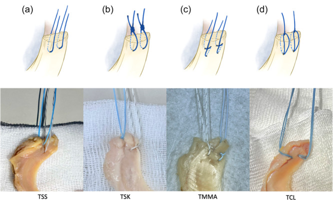
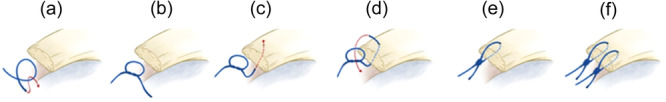
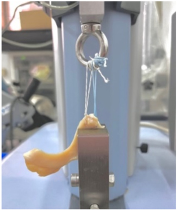
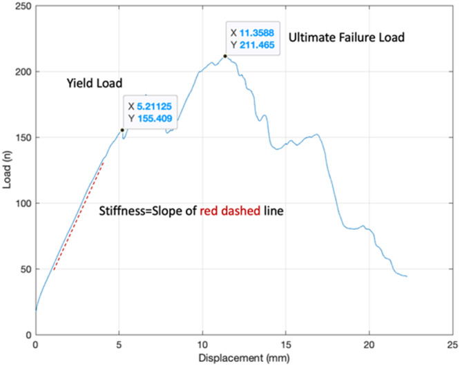
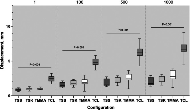
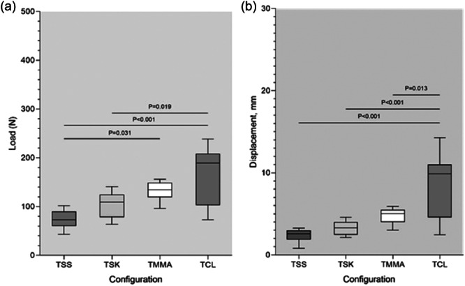
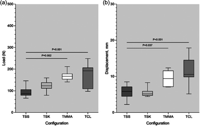
Method: Thirty-two fresh-frozen cadaveric menisci were randomly allocated to four meniscus-suture fixation constructs: Two simple-sutures (TSS), two slip-knot (TSK) sutures, two cinch-loop (TCL) sutures, and two modified Mason-Allen (TMMA) sutures. Cyclic loading from 5 to 20 N was conducted for 1000 cycles at 0.5 Hz, and then loaded to failure at 0.5 mm/s. Parametric data (displacement during cyclic loading, ultimate load, yield load, and displacement at failure) were analysed using a one-way analysis of variance (ANOVA), whereas nonparametric data (stiffness) were analysed using the Kruskal-Wallis test.
Results: After 1000 cycles, the TCL construct significantly displaced the most (mean ± SD, 6.78 ± 1.32 mm; p < 0.001), followed by the TMMA (2.83 ± 0.90 mm), TSK (2.33 ± 0.57 mm), and TSS (2.03 ± 0.62 mm) groups. On ultimate failure load, there was no significant difference between the TSK group (123.48 ± 27.24 N, p > 0.05) and the other three groups (TSS, 94.65 ± 25.33 N; TMMA, 168.38 ± 23.24 N; TCL, 170.54 ± 57.32 N); however, it exhibited the least displacement (5.53 ± 1.25 mm) which was significantly shorter than those of the TCL (11.82 ± 4.25 mm, p < 0.001) and TMMA (9.53 ± 2.18 mm, p = 0.03) constructs. No significant difference in stiffness was observed among the four meniscus-suture constructs.
Conclusion: The slip-knot technique has proven to be a simple, yet robust and stable meniscal root fixation option; moreover, it exhibited superiority over the more complex modified Mason-Allen suture construct in resisting displacement at the ultimate failure load.
Level of evidence: Not applicable.
Keywords: knee; meniscus root; pull‐out repair; root tear; slip‐knot.
© 2024 The Author(s). Journal of Experimental Orthopaedics published by John Wiley & Sons Ltd on behalf of European Society of Sports Traumatology, Knee Surgery and Arthroscopy.
Conflict of interest statement
The authors declare no conflict of interest.
Figures
Similar articles
-
Cyclic displacement after meniscal root repair fixation: a human biomechanical evaluation.Am J Sports Med. 2015 Apr;43(4):892-8. doi: 10.1177/0363546514562554. Epub 2015 Jan 2. Am J Sports Med. 2015. PMID: 25556220 Clinical Trial.
-
Biomechanical Performance of Transtibial Pull-Out Posterior Horn Medial Meniscus Root Repair Is Improved With Knotless Adjustable Suture Anchor-Based Fixation.Orthop J Sports Med. 2024 Apr 5;12(4):23259671241239575. doi: 10.1177/23259671241239575. eCollection 2024 Apr. Orthop J Sports Med. 2024. PMID: 38584990 Free PMC article.
-
Biomechanical Comparison of an All-Inside Meniscal Repair Device Construct Versus Pullout Sutures for Arthroscopic Transtibial Repair of Posterior Medial Meniscus Root Tears: A Matched-Pair Cadaveric Study.Orthop J Sports Med. 2021 Apr 22;9(4):23259671211000464. doi: 10.1177/23259671211000464. eCollection 2021 Apr. Orthop J Sports Med. 2021. PMID: 33997064 Free PMC article.
-
Biomechanical Properties of Posterior Meniscal Root Repairs: A Systematic Review.Arthroscopy. 2019 Jul;35(7):2189-2206.e2. doi: 10.1016/j.arthro.2019.01.018. Epub 2019 Apr 9. Arthroscopy. 2019. PMID: 30979628
-
Modified Mason-Allen vs Two Simple Stitch Fixation for Medial Meniscus Posterior Root Tears: A Systematic Review and Meta-analysis.Am J Sports Med. 2024 Jun;52(7):1877-1887. doi: 10.1177/03635465231190650. Epub 2024 Jan 23. Am J Sports Med. 2024. PMID: 38258492
References
-
- Bachmaier, S. , Krych, A.J. , Smith, P.A. , Feucht, M.J. , LaPrade, R.F. & Wijdicks, C.A. (2024) Biomechanical performance of transtibial pull‐out posterior horn medial meniscus root repair is improved with knotless adjustable suture anchor–based fixation. Orthopaedic Journal of Sports Medicine, 12(4), 23259671241239575. Available from: 10.1177/23259671241239575 - DOI - PMC - PubMed
-
- Cerminara, A.J. , LaPrade, C.M. , Smith, S.D. , Ellman, M.B. , Wijdicks, C.A. & LaPrade, R.F. (2014) Biomechanical evaluation of a transtibial pull‐out meniscal root repair: challenging the bungee effect. The American Journal of Sports Medicine, 42(12), 2988–2995. Available from: 10.1177/0363546514549447 - DOI - PubMed
LinkOut - more resources
Full Text Sources
Research Materials
