

Detective flow imaging versus contrast-enhanced EUS in solid pancreatic lesions
- PMID: 39318752
- PMCID: PMC11419480
- DOI: 10.1097/eus.0000000000000076
Detective flow imaging versus contrast-enhanced EUS in solid pancreatic lesions
Abstract
Background and objectives: Detective flow imaging EUS (DFI-EUS) is a new technology that detects fine vessels and low-flow velocity without contrast agents, used in real time during EUS, with a better resolution compared to usual technologies such as color Doppler and eFLOW. The aim of this study was to compare DFI-EUS with contrast-enhanced EUS (CE-EUS) for the evaluation of vascularization in solid pancreatic lesions.
Methods: We included patients who had a pancreatic mass visualized by EUS, with recorded images of their assessment in DFI-EUS and CE-EUS techniques and a histological diagnosis confirmed malignant tumors or a minimum of 1-year follow-up for benign lesions.
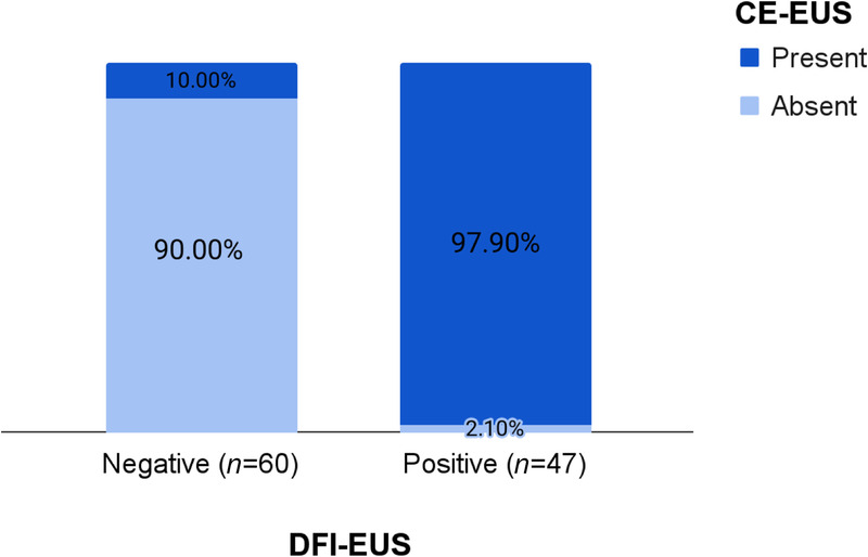
Results: Of the 107 patients included in this retrospective single-center study, the histological diagnosis revealed 69 cases (64.5%) of pancreatic adenocarcinoma, 18 cases (16.8%) of neuroendocrine tumors (NETs), and 10 cases (9.3%) of metastases from nonpancreatic cancers. A smaller proportion (9.4%) exhibited other lesions. As a result, the incidence of intralesional microvascularization was 43.9% with DFI-EUS and 48.6% with CE-EUS, indicating a positive correlation between the 2 techniques (P = 0.0001). Compared to CE-EUS, DFI-EUS exhibited sensitivity, specificity, positive predictive value (PPV), and negative predictive value (NPV) of 88.5%, 98.2%, 97.9%, and 90%, respectively, for the detection of intralesional vessels.
Conclusions: The novel technique DFI-EUS demonstrates a remarkable correlation with CE-EUS, exhibiting high sensitivity and specificity for the assessment of microvascularization in solid pancreatic lesions. This method eliminates the need for a contrast agent, thus carrying no risk of adverse effects.
Keywords: Contrast-enhanced EUS; Detective flow imagine EUS; Doppler EUS; Pancreatic cancer; Solid pancreatic lesions.
Copyright © 2024 The Author(s). Published by Wolters Kluwer Health, Inc on behalf of Scholar Media Publishing.
Conflict of interest statement
Marc Giovannini is a Founding Editor-in-Chief of the journal. The article was subjected to the standard procedures of the journal, with a review process independent of the editors and their research group. The other authors declare that they have no conflict of interest.
Figures


References
-
- Yamaguchi K Okusaka T Shimizu K, et al. . Clinical practice guidelines for pancreatic cancer 2016 from the Japan Pancreas Society: a synopsis. Pancreas 2017;46:595–604. - PubMed
-
- Giovannini M. Contrast-enhanced endoscopic ultrasound and elastosonoendoscopy. Best Pract Res Clin Gastroenterol 2009;23:767–779. - PubMed
LinkOut - more resources
Full Text Sources
Research Materials
