Review
. 2024 Aug 25;16(8):e67726.
doi: 10.7759/cureus.67726. eCollection 2024 Aug.

Evidence of Programmed Death-Ligand 1 Expression in a Highly Inflammatory Prostate: A Literature Review and Our Experience

Affiliations
Review

Evidence of Programmed Death-Ligand 1 Expression in a Highly Inflammatory Prostate: A Literature Review and Our Experience

Maria Koleva et al. Cureus. .

Abstract

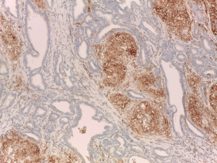
Chronic inflammation (CI), a common finding in the human prostate, is associated with the most frequent socially important prostate diseases: prostatitis, benign prostatic hyperplasia, and prostate adenocarcinoma. Programmed cell death protein 1 (PD-1) and its ligand (PD-L1) expression are induced on the surface of immune and epithelial cells of healthy and tumor tissues in response to various cytokines. Here, we provide a comprehensive review of the PD-1/PD-L1 pathway in the non- and peri-tumoral inflammatory prostate, focusing on the structure and expression of PD-L1 and the diverse biological functions of PD-L1 signaling in health, high-grade CI (National Institutes of Health, category IV prostatitis or histologic prostatitis), and immune-related diseases, including autoimmunity, tumor microenvironmental immunity, and immune privilege. This review explores the possible pathophysiological interpretations of clearly visible, selective, and strong PD-L1 expression in the immuno-inflammatory-induced and related, histologically distinct sites of this expression: the ductal lymphoepithelial lesions and prostatic granulomas.

Keywords: inflammation; lymphoepithelial lesions; pd-l1; prostate; prostatitis.


Conflict of interest statement

Conflicts of interest: In compliance with the ICMJE uniform disclosure form, all authors declare the following: Payment/services info: All authors have declared that no financial support was received from any organization for the submitted work. Financial relationships: All authors have declared that they have no financial relationships at present or within the previous three years with any organizations that might have an interest in the submitted work. Other relationships: All authors have declared that there are no other relationships or activities that could appear to have influenced the submitted work.

Figures

Figure 1
Figure 1. PD-L1 expression in HG-HP in serial sections.
The epithelium of the lymphoepithelial lesion showed selective and strong membranous immunoexpression of programmed death-ligand 1, not needing a special indication with arrows. Immunohistochemistry anti-programmed death-ligand 1, x200. HG: high grade; HP: histologic prostatitis; PD-L1: programmed death-ligand 1 Image credits: Dr. Dorian Dikov
Figure 2
Figure 2. Chronic HG prostatic inflammation.
Chronic high-grade (HG) prostatic inflammation with infiltration of lymphocytes affecting multiple prostatic ducts, associated with strongly eosinophilic epithelium, fully corresponding to programmed death-ligand 1-positive lymphoepithelial lesion from Figure 1; pronounced PD-L1 expression was also found in intra-luminal macrophages. Haematoxylin-eosin-saffron, x200. PD-L1: programmed death-ligand 1 Image credits: Dr. Dorian Dikov
Figure 3
Figure 3. PD-L1 expression in tonsillar crypts lymphoepithelium of adenoids (external positive control). Immunohistochemistry anti-PD-L1, x200.
PD-L1: programmed death-ligand 1 Image credits: Dr. Dorian Dikov
Figure 4
Figure 4. Foci of localized GP, haematoxylin-eosin-saffron, x100.
GP: granulomatous prostatitis Image credits: Dr. Dorian Dikov
Figure 5
Figure 5. PD-L1 expression in GP in serial sections.
Strong programmed death-ligand 1 (PD-L1) expression in the foci of localized granulomatous prostatitis. GP: granulomatous prostatitis Image credits: Dr. Dorian Dikov
Figure 6
Figure 6. The strong and selective PD-L1 immunoreactivity in HG-inflammatory prostate is easily noticeable even under scanning microscopic magnification.
The presence of four programmed death-ligand 1 (PD-L1)-positive stromal granulomas on the left side of the picture and one programmed death-ligand 1-positive lymphoepithelial lesion in the ductal epithelium in the right part (arrow). Immunohistochemistry anti-programmed death-ligand, x100. The strong and selective programmed death-ligand immunoreactivity in high-grade (HG) inflammatory prostate is easily noticeable even under scanning microscopic magnification: the presence of four programmed death-ligand-positive stromal granulomas on the left side of the picture and one programmed death-ligand-positive lymphoepithelial lesion in the ductal epithelium in the right part (arrow). Immunohistochemistry anti-programmed death-ligand 1, x100. Image credits: Dr. Dorian Dikov
Figure 7
Figure 7. General view of HG-HP with rupture of a ductal LEL and diffusion of the intraluminal substances among immune periductal cells. Haematoxylin-eosin-saffron; x50.
HG: high grade; HP: histologic prostatitis; LEL: lymphoepithelial lesions Image credits: Dr. Dorian Dikov
Figure 8
Figure 8. Damaged prostatic epithelium of ductal LEL in HG-HP.
Programmed death-ligand 1-positive lymphoepithelial lesion in close proximity with intraprostatic spermatozoa (on the left). HG: high grade; HP: histologic prostatitis; LEL: lymphoepithelial lesions Image credits: Dr. Dorian Dikov
Figure 9
Figure 9. Eroded LEL from intraluminal corpora amylacea, haematoxylin-eosin-saffron, x400.
LEL: lymphoepithelial lesions Image credits: Dr. Dorian Dikov
Figure 10
Figure 10. M2 intraprostatic macrophages in HG-HP, showing immunosuppressive phenotype CD163+.
HG: high grade; HP: histologic prostatitis Image credits: Dr. Dorian Dikov
Figure 11
Figure 11. M2 intraprostatic macrophages in HG-HP, PD-L1+.
HG: high grade; HP: histologic prostatitis; PD-L1: PD-L1: programmed death-ligand 1 Image credits: Dr. Dorian Dikov

References

    1. Inflammation in prostate carcinogenesis. De Marzo AM, Platz EA, Sutcliffe S, et al. Nat Rev Cancer. 2007;7:256–269. - PMC - PubMed
    1. The inflammatory microenvironment and microbiome in prostate cancer development. Sfanos KS, Yegnasubramanian S, Nelson WG, De Marzo AM. Nat Rev Urol. 2018;15:11–24. - PubMed
    1. Consensus development of a histopathological classification system for chronic prostatic inflammation. Nickel JC, True LD, Krieger JN, Berger RE, Boag AH, Young ID. BJU Int. 2001;87:797–805. - PubMed
    1. The prognostic role of immune checkpoint markers programmed cell death protein 1 (PD-1) and programmed death ligand 1 (PD-L1) in a large, multicenter prostate cancer cohort. Ness N, Andersen S, Khanehkenari MR, et al. Oncotarget. 2017;8:26789–26801. - PMC - PubMed
    1. Immunohistochemistry of immune checkpoint markers PD-1 and PD-L1 in prostate cancer. Sharma M, Yang Z, Miyamoto H. Medicine (Baltimore) 2019;98:0. - PMC - PubMed

LinkOut - more resources