Single-cell transcriptomic landscape reveals the role of intermediate monocytes in aneurysmal subarachnoid hemorrhage
- PMID: 39318997
- PMCID: PMC11420033
- DOI: 10.3389/fcell.2024.1401573
Single-cell transcriptomic landscape reveals the role of intermediate monocytes in aneurysmal subarachnoid hemorrhage
Abstract
Objective: Neuroinflammation is associated with brain injury and poor outcomes after aneurysmal subarachnoid hemorrhage (SAH). In this study, we performed single-cell RNA sequencing (scRNA-seq) to analyze monocytes and explore the mechanisms of neuroinflammation after SAH.
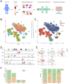
Methods: We recruited two male patients with SAH and collected paired cerebrospinal fluid (CSF) and peripheral blood (PB) samples from each patient. Mononuclear cells from the CSF and PB samples were sequenced using 10x Genomics scRNA-seq. Additionally, scRNA-seq data for CSF from eight healthy individuals were obtained from the Gene Expression Omnibus database, serving as healthy controls (HC). We employed various R packages to comprehensively study the heterogeneity of transcriptome and phenotype of monocytes, including monocyte subset identification, function pathways, development and differentiation, and communication interaction.
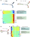
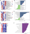
Results: (1) A total of 17,242 cells were obtained in this study, including 7,224 cells from CSF and 10,018 cells from PB, mainly identified as monocytes, T cells, B cells, and NK cells. (2) Monocytes were divided into three subsets based on the expression of CD14 and CD16: classical monocytes (CM), intermediate monocytes (IM), and nonclassical monocytes (NCM). Differentially expressed gene modules regulated the differentiation and biological function in monocyte subsets. (3) Compared with healthy controls, both the toll-like receptor (TLR) and nod-like receptor (NLR) pathways were significantly activated and upregulated in IM from CSF after SAH. The biological processes related to neuroinflammation, such as leukocyte migration and immune response regulation, were also enriched in IM. These findings revealed that IM may play a key role in neuroinflammation by mediating the TLR and NLR pathways after SAH.
Interpretation: In conclusion, we establish a single-cell transcriptomic landscape of immune cells and uncover the heterogeneity of monocyte subsets in SAH. These findings offer new insights into the underlying mechanisms of neuroinflammation and therapeutic targets for SAH.
Keywords: ScRNA-seq; TLR; aneurysmal subarachnoid hemorrhage; monocytes; neuroinflammation.
Copyright © 2024 Meng, Su, Ye, Xie, Liu and Qin.
Conflict of interest statement
The authors declare that the research was conducted in the absence of any commercial or financial relationships that could be construed as a potential conflict of interest.
Figures


Similar articles
-
Immune cells subpopulations in cerebrospinal fluid and peripheral blood of patients with Aneurysmal Subarachnoid Hemorrhage.Springerplus. 2015 Apr 23;4:195. doi: 10.1186/s40064-015-0970-2. eCollection 2015. Springerplus. 2015. PMID: 25977890 Free PMC article.
-
Single-cell RNA sequencing reveals that an imbalance in monocyte subsets rather than changes in gene expression patterns is a feature of postmenopausal osteoporosis.J Bone Miner Res. 2024 Aug 5;39(7):980-993. doi: 10.1093/jbmr/zjae065. J Bone Miner Res. 2024. PMID: 38652170
-
The significance of CD16+ monocytes in the occurrence and development of chronic thromboembolic pulmonary hypertension: insights from single-cell RNA sequencing.Front Immunol. 2024 Aug 13;15:1446710. doi: 10.3389/fimmu.2024.1446710. eCollection 2024. Front Immunol. 2024. PMID: 39192976 Free PMC article.
-
Interleukin 6 and Aneurysmal Subarachnoid Hemorrhage. A Narrative Review.Int J Mol Sci. 2021 Apr 16;22(8):4133. doi: 10.3390/ijms22084133. Int J Mol Sci. 2021. PMID: 33923626 Free PMC article. Review.
-
Toll-like receptor 4 as a possible therapeutic target for delayed brain injuries after aneurysmal subarachnoid hemorrhage.Neural Regen Res. 2017 Feb;12(2):193-196. doi: 10.4103/1673-5374.200795. Neural Regen Res. 2017. PMID: 28400792 Free PMC article. Review.
References
LinkOut - more resources
Full Text Sources
Research Materials

