Cardiogenic shock in phaeochromocytoma multisystem crisis: a case report
- PMID: 39319178
- PMCID: PMC11420679
- DOI: 10.1093/ehjcr/ytae463
Cardiogenic shock in phaeochromocytoma multisystem crisis: a case report
Abstract
Background: Phaeochromocytoma multisystem crisis (PMC) is characterized by labile blood pressures (extremes of hypo- and/or hypertension) and multiorgan failure as a result of catecholamine excess. Initial stabilization requires pharmacological and/or mechanical circulatory support, followed by the institution of antihypertensives to correct the underlying pathophysiology.
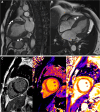
Case summary: A previously well 40-year-old male developed a sudden onset of breathlessness. On presentation, he was in shock with multiorgan failure. He required intubation, mechanical ventilation, dual inotropic support, and renal replacement therapy. Bedside echocardiogram showed a severely impaired left ventricular ejection fraction (LVEF) of 25%. Coronary angiography revealed normal coronary arteries. In view of raised inflammatory markers and transaminitis, a computed tomography abdomen/pelvis was performed. An incidental left adrenal mass was found. Further work-ups revealed raised plasma metanephrine and normetanephrine, 24-h urine epinephrine, and norepinephrine. A cardiac magnetic resonance (CMR) showed myocardial inflammation and reverse Takotsubo pattern of regional wall motion abnormality (RWMA). The diagnosis of cardiogenic shock and stress cardiomyopathy secondary to PMC was made. He was subsequently initiated on α- and β-blockers and goal-directed medical therapy for heart failure. A 68Ga-DOTATATE scan showed avid tracer uptake of the left phaeochromocytoma. An interval CMR 3 weeks from presentation showed near normalization of the LVEF and RWMA. He underwent a successful laparoscopic left adrenalectomy and was antihypertensive-free since.
Discussion: The clinical suspicion for PMC as the cause of cardiogenic shock requires astute clinical judgement, while the management requires an understanding of the underlying pathophysiology that calls for multidisciplinary inputs.
Keywords: Case report; Catecholamines; Phaeochromocytoma; Phaeochromocytoma multisystem crisis.
© The Author(s) 2024. Published by Oxford University Press on behalf of the European Society of Cardiology.
Conflict of interest statement
Conflict of interest: The authors declared no conflict of interest.
Figures


References
-
- Newell KA, Prinz RA, Pickleman J, Braithwaite S, Brooks M, Karson TH, et al. . Pheochromocytoma multisystem crisis a surgical emergency. Arch Surg 1988;123:956–959. - PubMed
-
- Ando Y, Ono Y, Sano A, Fujita N, Ono S, Tanaka Y. Clinical characteristics and outcomes of pheochromocytoma crisis: a literature review of 200 cases. J Endocrinol Invest 2022;45:2313–2328. - PubMed
-
- Brouwers FM, Eisenhofer G, Lenders JWM, Pacak K. Emergencies caused by pheochromocytoma, neuroblastoma, or ganglioneuroma. Endocrinol Metab Clin North Am 2006;35:699–724. - PubMed
-
- Prejbisz A, Lenders JWM, Eisenhofer G, Januszewicz A. Cardiovascular manifestations of phaeochromocytoma. J Hypertens 2011;29:2049–2060. - PubMed
-
- Gruber LM, Hartman RP, Thompson GB, McKenzie TJ, Lyden ML, Dy BM, et al. . Pheochromocytoma characteristics and behavior differ depending on method of discovery. J Clin Endocrinol Metab 2019;104:1386–1393. - PubMed
Publication types
LinkOut - more resources
Full Text Sources
