. 2024 Sep 4;27(10):110883.
doi: 10.1016/j.isci.2024.110883. eCollection 2024 Oct 18.

Regenerative potential of mouse neonatal intervertebral disc depends on collagen crosslink density

Affiliations

Regenerative potential of mouse neonatal intervertebral disc depends on collagen crosslink density

Danielle N D'Erminio et al. iScience. .

Abstract

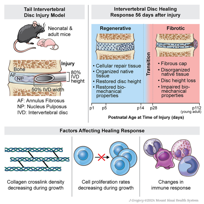
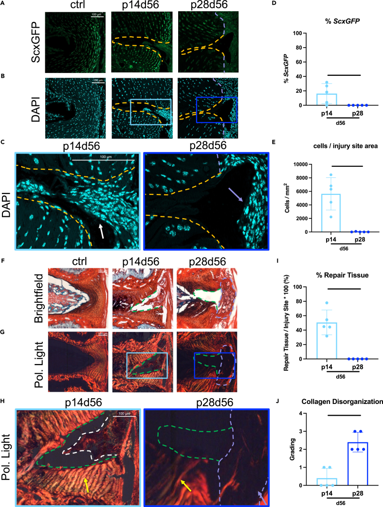
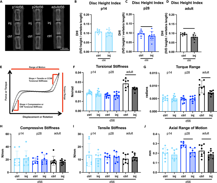
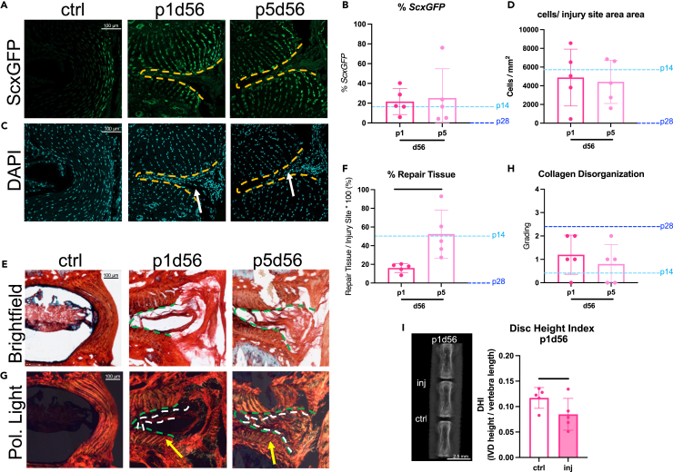
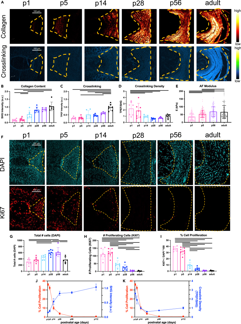
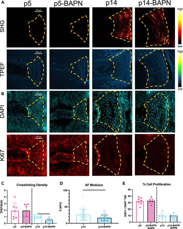
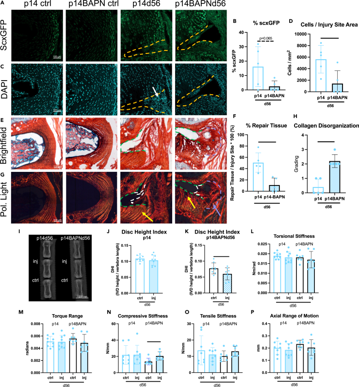
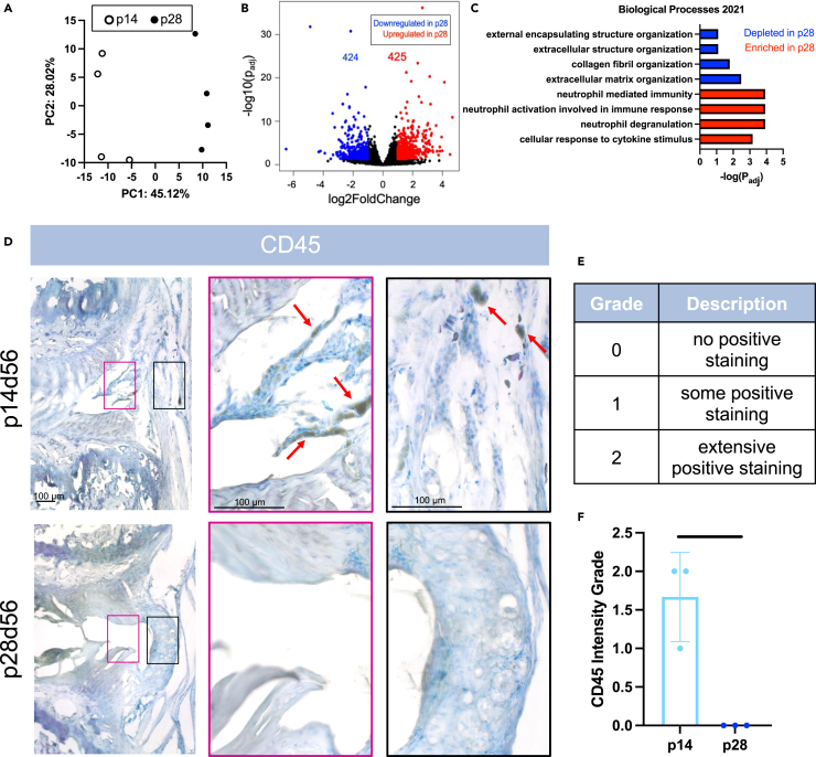
Intervertebral disc (IVD) defects heal poorly and can cause back pain and disability. We identified that IVD herniation injury heals regeneratively in neonatal mice until postnatal day 14 (p14) and shifts to fibrotic healing by p28. This age coincides with the shift in expansive IVD growth from cell proliferation to matrix elaboration, implicating collagen crosslinking. β-aminopropionitrile treatment reduced IVD crosslinking and caused fibrotic healing without affecting cell proliferation. Bulk sequencing on naive IVDs was depleted for matrix structural organization from p14 to p28 to validate the importance of crosslinking in regenerative healing. We conclude that matrix changes are key drivers in the shift to fibrotic healing, and a stably crosslinked matrix is needed for IVD regeneration.

Keywords: Molecular biology; Omics; Tissue Engineering; Transcriptomics.


Conflict of interest statement

The authors declare no competing interests.

Figures

None
Graphical abstract
Figure 1
Figure 1
Injured p14 IVDs heal regeneratively with highly cellular matrix deposition throughout the injury site and organized AF layers adjacent to the injury while injured p28 IVDs heal with fibrosis (A–J) Representative (A) ScxGFP and (B) DAPI images of AF controls and injury sites of p14 and p28 mice 56 days after injury with (C) magnified images of injury sites with DAPI. Quantification of (D) % cells expressing ScxGFP and (E) total number of cells within injury sites. Picrosirius Red Alcian blue images of AF controls and injury sites of p14d56 and p28d56 mice under (F) brightfield and (G) polarized light with (H) magnified images of injury sites of p14d56 and p28d56. Quantification of (I) % repair tissue within the injury site and (J) collagen disorganization score. Scale bars = 100 μm. Dashed lines outline injury sites (yellow or green), fibrous caps (purple), and repair tissue (white). Arrows point toward cellular repair tissue (white), fibrous caps (purple), and adjacent AF structures near injury sites (yellow). Biological n = 5/age, error bars = SD. Student’s t test with p < 0.05 determined significance, depicted in graphs as a horizontal black line.
Figure 2
Figure 2
Injured p14 IVDs heal with restored disc height index (DHI) and biomechanical properties by d56 while injured p28 IVDs and adult IVDs do not (A) digital X-rays used to calculate DHI in (B) p14, (C) p28, and (D) adult mice (biological n = 8–9/age) 56 days after injury. Scale bars represent 2.5 mm. (E) Representative loading curve for samples used to determine torsional (F and G) and (H–J) axial biomechanical parameters. Error bars = SD. Student’s t test with p < 0.05 determined significant differences between ctrl and inj for each age, depicted in graphs as a horizontal black line.
Figure 3
Figure 3
Injured p1 and p5 IVDs heal near-regeneratively suggesting neonatal IVD regeneration does not recapitulate the AF lamellar structure after a severe herniation-type injury at any post-natal age (A and B) Representative (A) ScxGFP and (B) DAPI images of AF controls and injury sites of p1 and p5 mice (biological n = 5/age) 56 days after injury. (C and D) Quantification of (C) % cells expressing ScxGFP and (D) total number of cells within injury sites. (E and F) Representative Picrosirius Red Alcian blue images of AF controls and injury sites of p1d56 and p5d56 mice under (E) brightfield and (F) polarized light. (G and H) Quantification of (G) % repair tissue within the injury site and (H) collagen disorganization score. Scale bars = 100 μm. Quantifications are compared to p14 and p28 levels marked by light and dark blue dotted lines respectively. Outlines indicate injury sites (yellow/green) and repair tissue (white). (I) Arrows point toward cellular repair tissue (white) and adjacent AF structure near injury sites (yellow) (I) Digital X-rays are used to calculate DHI in p1 injured and control IVDs. Scale bars represent 2.5 mm. Error bars = SD. Student’s t test with p < 0.05 determined significance, depicted in graphs as a horizontal black line.
Figure 4
Figure 4
Postnatal AF growth involves rapidly increased collagen accumulation and AF modulus with reduced crosslinking density and AF cell proliferation (A) Representative IVD images (biological n = 6–9/age) under second harmonic generation (SHG, excitation 910 nm) depicting collagen content, and two photon excitation fluorescence (TPEF, excitation 720nm) depicting crosslinking. Scale bars = 100 μm. (B–D) Quantification of (B) collagen content (C) crosslinking and (D) crosslinking density (TPEF/SHG). (E) Quantification of AF elastic modulus, E. (F–I) Representative AF images (n = 6–9/age) of DAPI and Ki67 (cell proliferation) Quantification of (G) total number of AF cells (H) number of proliferating cells, and (I) % cell proliferation. (J) Inverse relationship between % AF cell proliferation (Ki67/DAPI) and collagen content (SHG). (K) Proportional relationship between % AF cell proliferation (Ki67/DAPI) and crosslink density (TPEF/SHG). Error bars = SD. Effect of age was evaluated by one way ANOVA with Tukey post-hoc comparisons. p < 0.05 is considered significant and indicated in graphs as a horizontal black line.
Figure 5
Figure 5
Inhibiting crosslinking accumulation with BAPN reduced AF crosslinking density and modulus at p14 without affecting % cell proliferation BAPN was administered through drinking water until embryonic day 5.5 until p5 or p14 (biological n = 6–9/age). (A and B) Representative (A) multiphoton images of SHG (collagen content) and TPEF (collagen crosslinking) and (B) fluorescent images of total cells (DAPI) and proliferating cells (Ki67) in control and BAPN-treated p5 and p14 AF tissue. Scale bars = 100 μm. (C–E) Quantification of (C) collagen crosslinking density (TPEF/SHG) (D) AF tissue elastic modulus, and (E) % cell proliferation. Dashed lines outline AF region (yellow). Error bars = SD. Student’s t test with p < 0.05 determined significance, depicted in graphs as a horizontal black line. p5 and p14 mice were the same as in the growth studies as all groups were processed and analyzed together.
Figure 6
Figure 6
BAPN treatment shifted p14 regenerative healing to fibrotic healing with little matrix/cell deposition, fibrotic caps, reduced DHI, and increased compressive stiffness BAPN-treated mice were injured at p14 and evaluated 56 days after injury (n = 5). (A–H) Representative fluorescent images of (A) ScxGFP (AF cells) and (B) DAPI (total number of cells) are used to quantify (C) cells/injury site area and (D) % with ScxGFP. Representative PRAB (E) brightfield and (F) polarized light images are used to quantify (G) % Repair Tissue and (H) Collagen Disorganization grade. Scale bars = 100 μm. Dashed lines outlines injury sites (yellow or green), fibrous caps (purple), and repair tissue (white). Arrows point toward cellular repair tissue (white), fibrous caps (purple), and adjacent AF structures near injury sites (yellow). (I) Digital X-rays were used to calculate DHI in (J) p14 and (K) p14BAPN mice 56 days after injury (biological n = 6–9/age). Torsional (L and M) and (N and P) axial biomechanical parameters. Error bars = SD. Student’s t test with p < 0.05 determined significance, depicted in graphs as a horizontal black line. p14d56 mice were the same as in the regeneration window studies as all groups were processed and analyzed together.
Figure 7
Figure 7
p28 AF has depleted structural organization pathways, enriched immune-related pathways, and fewer CD45-immune cells at the injury site compared to p14 (A–C) Bulk sequencing of p14 and p28 AF tissue (n = 4 samples/age) revealed (A) PCA plot highlighting p14 and p28 gene expression profiles are distinct with (B) volcano plot depicting the number of differentially expressed genes (DEGs) and (C) GO-Terms found using Enrichr highlighting the most significant enriched and depleted pathways. (D) Immunohistochemical staining of leukocyte cell marker CD45 using a peroxidase/DAB secondary kit with a TOL Blue counterstain on p14 and p28 IVDs (n = 3/age) 56 days after injury. Arrows (red) point toward representative positive cells (black). Pink and black insets highlight areas within injury sites within the boundary of the IVD (pink) and on the outer periphery (black). (E) Scale bars = 100 μm (E) CD45+ staining semi-quantification grading scale. (F) CD45+ grading for p14 and p28 IVDs 56 days after injury. Error bars = SD. Student’s t test with p < 0.05 determined significance, depicted in graphs as a horizontal black line.

Similar articles

Cited by

References

    1. Adams M.A., Roughley P.J. What is intervertebral disc degeneration, and what causes it? Spine. 2006;31:2151–2161. doi: 10.1097/01.brs.0000231761.73859.2c. - DOI - PubMed
    1. Mohanty S., Dahia C.L. Defects in intervertebral disc and spine during development, degeneration, and pain: New research directions for disc regeneration and therapy. Wiley Interdiscip. Rev. Dev. Biol. 2019;8 doi: 10.1002/wdev.343. - DOI - PMC - PubMed
    1. Hartvigsen J., Hancock M.J., Kongsted A., Louw Q., Ferreira M.L., Genevay S., Hoy D., Karppinen J., Pransky G., Sieper J., et al. What low back pain is and why we need to pay attention. Lancet. 2018;391:2356–2367. doi: 10.1016/S0140-6736(18)30480-X. - DOI - PubMed
    1. Dieleman J.L., Cao J., Chapin A., Chen C., Li Z., Liu A., Horst C., Kaldjian A., Matyasz T., Scott K.W., et al. US Health Care Spending by Payer and Health Condition, 1996-2016. JAMA. 2020;323:863–884. doi: 10.1001/jama.2020.0734. - DOI - PMC - PubMed
    1. Iatridis J.C., Nicoll S.B., Michalek A.J., Walter B.A., Gupta M.S. Role of biomechanics in intervertebral disc degeneration and regenerative therapies: what needs repairing in the disc and what are promising biomaterials for its repair? Spine J. 2013;13:243–262. doi: 10.1016/j.spinee.2012.12.002. - DOI - PMC - PubMed