. 2024 Nov 11;52(20):12712-12726.
doi: 10.1093/nar/gkae826.

Phylogenomic instructed target analysis reveals ELAV complex binding to multiple optimally spaced U-rich motifs

Affiliations

Phylogenomic instructed target analysis reveals ELAV complex binding to multiple optimally spaced U-rich motifs

David W J McQuarrie et al. Nucleic Acids Res. .

Abstract

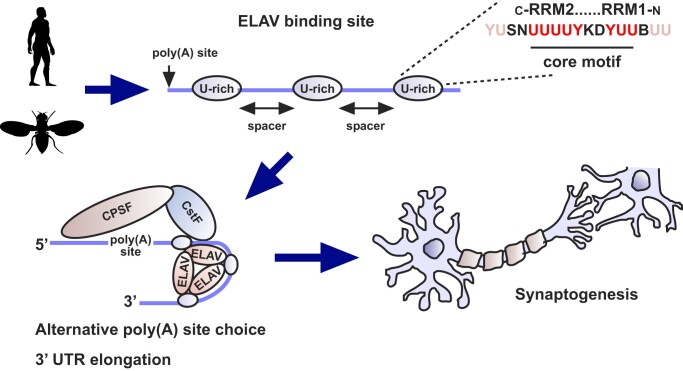
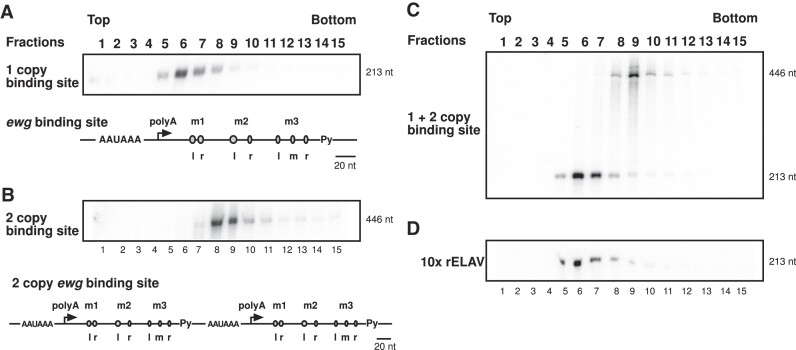
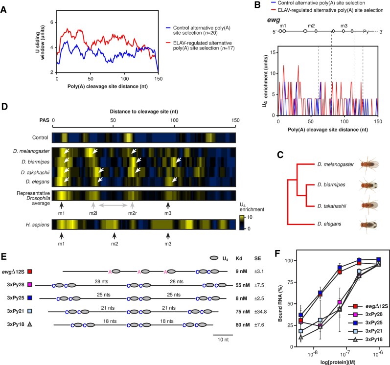
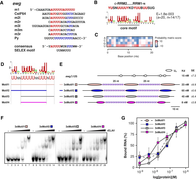
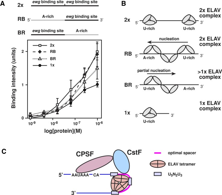
ELAV/Hu RNA-binding proteins are gene-specific regulators of alternative pre-mRNA processing. ELAV/Hu family proteins bind to short AU-rich motifs which are abundant in pre-mRNA, making it unclear how they achieve gene specificity. ELAV/Hu proteins multimerize, but how multimerization contributes to decode degenerate sequence environments remains uncertain. Here, we show that ELAV forms a saturable complex on extended RNA. Through phylogenomic instructed target analysis we identify the core binding motif U5N2U3, which is repeated in an extended binding site. Optimally spaced short U5N2U3 binding motifs are key for high-affinity binding in this minimal binding element. Binding strength correlates with ELAV-regulated alternative poly(A) site choice, which is physiologically relevant through regulation of the major ELAV target ewg in determining synapse numbers. We further identify a stem-loop secondary structure in the ewg binding site unwound upon ELAV binding at three distal U motifs. Base-pairing of U motifs prevents ELAV binding, but N6-methyladenosine (m6A) has little effect. Further, stem-loops are enriched in ELAV-regulated poly(A) sites. Additionally, ELAV can nucleate preferentially from 3' to 5'. Hence, we identify a decisive mechanism for ELAV complex formation, addressing a fundamental gap in understanding how ELAV/Hu family proteins decode degenerate sequence spaces for gene-specific mRNA processing.


Figures

Graphical Abstract
Graphical Abstract
Figure 1.
Figure 1.
ELAV forms a saturable complex on ewg RNA. (A–D) Glycerol gradient centrifugation fractions from top to bottom of recombinant ELAV with (A) 1 copy ELAV binding site RNA from ewg, (B) 2 copy ELAV binding site RNA from ewg, (C) 1 and 2 mixed copy ELAV binding site RNA from ewg and (D) 1 copy ELAV binding site RNA from ewg with a 10-fold increased ELAV concentration. After RNA extraction, fractions were run on 8% denaturing gels. 1 and 2 copy binding site RNAs ran in distinctly different positions (A–C), while increasing the concentration of ELAV had no effect on complex size (D). As detailed in A, the ELAV binding site in ewg harbours three U-rich tandem motifs termed m1, m2 and a triple U-motif in m3. RNA substrates are depicted in (A) and (B) with m1, m2, m3 and respectively left (l), right (r) and middle (m) U-rich sequences highlighted, described in Soller and White (21).
Figure 2.
Figure 2.
Spaced U-rich motif architecture is evolutionarily conserved between Drosophila and humans. (A and B) Comparative U and U4 enrichment sliding window scores calculated for 150 nucleotides downstream of control (blue) and ELAV-regulated (red) alternative poly(A) site selection gene cleavage sites in D. melanogaster. Positions of relevant high-affinity ewg ELAV binding sites are indicated spatially in schematics above (C). (C) Phylogeny of the Drosophila species analysed (D. melanogaster, D. biarmipes, D. takahashii and D. elegans). (D) U4 enrichment in control and ELAV-regulated alternative poly(A) site selection genes compared between D. melanogaster, D. biarmipes, D. takahashii, D. elegans and H. sapiens for the 150-nucleotide region downstream of poly(A) cleavage sites. Enriched motif positions are indicated with black and grey arrows labelled as m1, m2 (m2l: left and m2r: right) and m3. Predominant motif positions were calculated based on the highest three spatial conservation scores between the four motifs within the Drosophila average range (Supplementary Dataset 1). (E and F) Schematic depiction of the ewg and synthetic RNAs used for EMSA analysis of motif spacing effects on ELAV binding affinity (E). The ewgΔ12S control RNA (only ewg, no vector sequence as in ewgΔ12 (21)) was used as a high-affinity positive control. EMSA results were quantified and plotted (F). The average and standard error at each concentration were calculated from at least two biological replicates (E). Kd values and standard deviation are shown in (F).
Figure 3.
Figure 3.
ELAV requires a conserved, defined U-rich motif for high-affinity binding. (A) Alignment of consecutive AU4-6 motifs from the ewg ELAV binding site. AU4-6 motifs are in red and the PyU2 motif is blue. The CstF64 binding site likely represents a suboptimal AU4-6 motif. A putative consensus sequence for RRM1 and RRM2 is indicated at the bottom. Sequence labels are in reference to ewg U-rich sequence positions, shown in Figure 1A and B, and detailed in Soller and White, 2005 (21). (B and C) Comparative de novo extended motif found from alignment of the 200-nucleotide poly(A) cleavage site downstream regions of 17 ELAV-regulated gene targets (see Figure 7B for the full list of genes). A putative consensus sequence for RRM1 and RRM2 is indicated at the top. (D and E) Consensus motif analysis of the analysed ELAV targets with schematic depiction of deletion mutagenesis motifs generated to test core components of the extended ELAV binding site, labelled Motif1 to 4 (D). RNA substrate schematics are indicated, incorporating optimal spacing and unpairing spacer sequences (E). The ewgΔ12S control RNA was used as a high-affinity positive control. (F and G) EMSA analysis of the synthetic RNA substrates (F) and quantification of ELAV complex formation (G). The average and standard error at each concentration were calculated from at least three biological replicates (G). Kd values and standard deviation are shown in (E).
Figure 4.
Figure 4.
ELAV-regulated alternative poly(A) site selection depends on a high-affinity binding site. (A) U4 enrichment sliding window scores calculated for 100 nucleotides downstream of ELAV-regulated genes with alternative poly(A) site selection. Genes are separated into three categories: strong (>4-fold change), intermediate (<4-fold, >2-fold change) and weak (<2-fold change) based on their last exon usage fold change between wild type and ELAV null mutants (49,50). Motif positions relative to those shown in Figure 2D are labelled (m1, m2, m3). (B–E) Analysis of sequence change accumulation using phyloP scores (B) and evolutionary conservation (D and E) between the closely related melanogaster subgroup, D. biarmipes (suzukii subgroup), D. takahashii (takahashii subgroup) and D. elegans (elegans subgroup) (C) for each nucleotide type (A, C, G, U) in each fold change category. Statistically significant differences from chi squared tests are indicated by asterisks (Bonferroni corrected *P ≤ 0.05, **P ≤ 0.01, ****P ≤ 0.0001). (F and G) EMSA analysis of the synthetic RNA substrates made for 5PtaseI, CG4662, Teh2 and Ect4 (F) and quantification of ELAV complex formation (G). The average and standard error at each concentration were calculated from at least two biological replicates (G). Kd values and standard deviation are shown in (G).
Figure 5.
Figure 5.
ELAV binding is required for synapse formation. (A) Schematic of the tcgER ewg transgenic rescue construct. EWG is expressed in the nervous system from a chimeric cDNA/genomic construct. The pA2 ELAV binding site is detailed with description of U-rich motifs m1, m2 and m3. Mutants were generated for m1-3, m1, m1-2, m2-3, and m3 as described in Soller and White (2005). (B, C) Representative images (B) and quantification (C) of NMJs from muscle 13 synapses for the analysed genotypes in an ewg null background (ewgl1). Motor neurons are stained with anti-HRP (magenta) and synaptic boutons with anti-NC82 (green, white in the merged picture). Statistically significant differences from ANOVA with Tukey's multiple comparisons correction are indicated by asterisks (*P ≤ 0.05, **P ≤ 0.01, ***P ≤ 0.001 adjusted P values). The scale bar (B) is 10 μm.
Figure 6.
Figure 6.
The minimal ELAV binding site in ewg contains a stem–loop structure which is unwound upon binding. (A) Enzymatic probing of ewg RNA pA2-IΔ12 for secondary structure and RNase footprinting of ELAV. (A) Enzymatic probing of ewg RNA pA2-IΔ12. Sensitivity of 5′ 32P labelled substrate RNA (100 pM) was tested for two concentrations of RNases A, T1, T2, V1 and micrococcal nuclease (MN) as indicated on top. L: single nucleotide ladder. RNase A cleaves preferentially after single stranded pyrimidines but can also cleave in helical regions. RNase T1 cleaves after single stranded G residues. RNase T2 cleaves single stranded regions with preference for A residues. Micrococcal nuclease cleaves single stranded regions. RNase V1 cleaves double stranded regions but can also cleave helical single strands. (B) Summary of enzymatic probing results from at least three experiments for each RNase. Secondary structure predictions were obtained with mfold and adjusted to experimental data. Nucleotides sensitive to hydrolysis are bold (nucleotides 47–55). The polypyrimidine tract (Py) is indicated by a dotted line. RNase cut strength is shown as (+) very weak, + weak, ++ moderate, and +++ strong. (C) RNase footprinting of ewg RNA pA2-IΔ12 in the presence of increasing amounts of ELAV (0.2, 0.8 and 3.2 μM) or in the absence of ELAV (–). Sequence landmarks (polyU motifs: m3l, m3m, m3r and Py) and protected sites (black bars) are indicated on the right. The black line to the left of the micrococcal nuclease series (MN, lanes 14–17) indicates nucleotides 25–46 which become nuclease-sensitive at the lowest ELAV concentration (0.2 μM). L: single nucleotide ladder. (D) Schematic summary of footprinting results with 0.2 μM ELAV in the context of pA2-IΔ12 secondary structure as determined in (A) and (B). Strongly protected nucleotides are circled and weak protection is indicated with broken circles. polyU motifs are bold (m3l, m3m, m3r and Py). The polypyrimidine tract is also indicated by a dotted line.
Figure 7.
Figure 7.
ELAV-regulated alternative poly(A) site selection gene targets preferentially contain stem–loop secondary structures. (A) Schematic of the downstream cleavage site region analysed for secondary structures in ELAV-regulated alternative poly(A) site selection genes, with ewg as an example. (B and C) Quantification of distinct ewg-like stem–loop structures in 200-nucleotide 5′ and 3′ regions of the control group and in ELAV targets for individual targets and as an overall percentage. Statistically significant differences from non-parametric chi-squared tests are indicated by asterisks (***P ≤ 0.001, ****P ≤ 0.0001 following Bonferroni correction). (D) Quantification of complimentary base pair changes in poly(A) cleavage site downstream regions of control and ELAV regulated genes. Complementary changes were classified as a nucleotide replacement required for stem–loop pairing between two species which maintained pairing. Statistically significant differences from non-parametric chi-squared tests are indicated by asterisks (***P ≤ 0.001 following Bonferroni correction). (E) Positions of stem–loops from ELAV target genes mapped onto the ewg ELAV binding site.
Figure 8.
Figure 8.
ELAV nucleates from a high-affinity binding site preferentially from 3′ to 5′. (A) Quantification of ELAV-RNA binding intensity (arbitrary units) for each of the cross-linked RNA variants run on denaturing gels. The 2× and 1× RNAs form regular complexes around the sizes of 1× and 2× ELAV complexes. The RB RNA (antisense site in the 5′ and a U-rich binding site in the 3′) formed a full 2× sized complex at high concentrations. The BR RNA (U-rich binding site in the 5′ and an antisense site in the 3′) formed an intermediary sized complex between 1× and 2× in size. (B) Schematic of the results from quantification of cross-linking gels showing the predicted mode by which ELAV forms larger complexes on single copy binding site RNA through nucleation with a directional 3′ to 5′ preference (shown through BR and RB) preference. (C) Schematic of CPSF, CstF, and ELAV binding to an ELAV target pre-mRNA with optimally spaced U-rich motifs. CPSF and CstF co-operative binding with ELAV in the ewg regulated poly(A) site selection could limit ELAV spreading via nucleation.

Similar articles

Cited by

References

    1. Matlin A.J., Clark F., Smith C.W.. Understanding alternative splicing: towards a cellular code. Nat. Rev. Mol. Cell Biol. 2005; 6:386–398. - PubMed
    1. Rogalska M.E., Vivori C., Valcárcel J.. Regulation of pre-mRNA splicing: roles in physiology and disease, and therapeutic prospects. Nat. Rev. Genet. 2023; 24:251–269. - PubMed
    1. Soller M. Pre-messenger RNA processing and its regulation: a genomic perspective. Cell. Mol. Life Sci. 2006; 63:796–819. - PMC - PubMed
    1. Tian B., Manley J.L.. Alternative polyadenylation of mRNA precursors. Nat. Rev. Mol. Cell Biol. 2017; 18:18–30. - PMC - PubMed
    1. Vuong C.K., Black D.L., Zheng S.. The neurogenetics of alternative splicing. Nat. Rev. Neurosci. 2016; 17:265–281. - PMC - PubMed

LinkOut - more resources