

Bleaching as a complement to fluoride-enhanced remineralization or resin infiltration in masking white spot lesions
- PMID: 39319903
- PMCID: PMC11464075
- DOI: 10.1590/1678-7757-2024-0097
Bleaching as a complement to fluoride-enhanced remineralization or resin infiltration in masking white spot lesions
Abstract
Objective: There are many suitable strategies for addressing caries, which is an ongoing worldwide problem. Although white spot lesions (WSLs) can be either remineralized naturally or treated with non- or micro-invasive strategies, their whitish and opaque appearance may persist. To evaluate the effects of tooth bleaching as a complement to fluoride-enhanced remineralization or resin infiltration in masking WSLs, as well as in enamel surface roughness relative to that of the adjacent enamel.
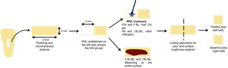
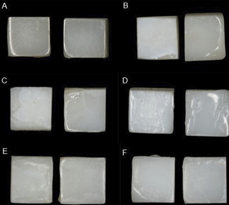
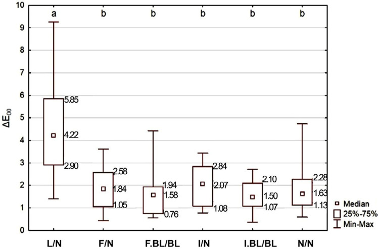
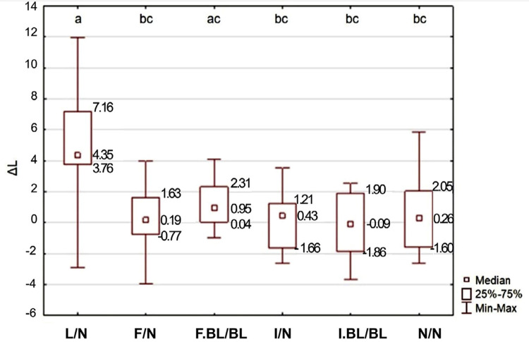
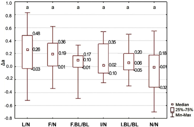
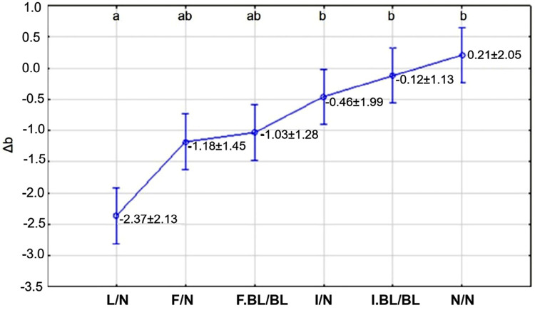
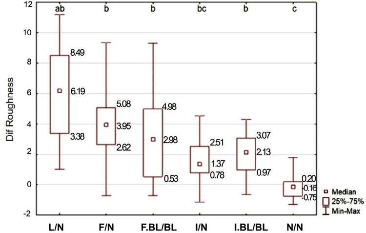
Methodology: Flattened rectangular bovine enamel fragments (6×3×~2.9 mm length, width and thickness) were divided into six groups (L/N, F/N, F.BL/BL, I/N, I.BL/BL, N/N; n=15). Treatments applied to the 3×3 mm left half included: L (Lesion) - WSL simulation with 50 mM acetate buffer, 96 hours, 37ºC; F (Fluoride) - WSL treatment with 2% NaF neutral gel, 1x/week, 8 weeks; I (Infiltration) - WSL treatment with H3PO4 37%/10 s; Icon®-Dry/30 s; Icon®-Infiltrant/3 min+1 min; N (Nothing) - sound enamel/control. Treatments applied to both halves after F and I included: BL (Bleaching) - Opalescence Boost 40%, 3×/20 min each; N (Nothing) - control. The differences in color (ΔE00, ΔL, Δa, Δb) and surface roughness (ΔRa) between the left and right halves were measured. Kruskal-Wallis/post-hoc tests were applied to ΔE00, ΔL, Δa and ΔRa, and 1-way ANOVA/Tukey tests to Δb (α=0.05).
Results: The factor under study significantly influenced ΔE00 (p=0.0001), ΔL (p=0.0024), Δb (p=0.0015), and ΔRa (p<0.001), but not Δa (p=0.1592). Both fluoride-enhanced remineralization and resin infiltration were able to mask WSL, regardless of subsequent bleaching. However, when bleaching was performed, ΔE00 median values did not exceed the acceptability threshold for color difference. Only resin infiltration reduced ΔRa between WSL and the adjacent enamel.
Conclusions: Both remineralization and infiltration, particularly if complemented by bleaching, fostered satisfactory esthetic results. Only infiltration without bleaching led to really good results in surface roughness.
Conflict of interest statement
Figures
Similar articles
-
Colour improvement and stability of white spot lesions following infiltration, micro-abrasion, or fluoride treatments in vitro.Eur J Orthod. 2014 Oct;36(5):595-602. doi: 10.1093/ejo/cjt095. Epub 2014 Jan 2. Eur J Orthod. 2014. PMID: 24385411
-
Aesthetic impact of resin infiltration and its mechanical effect on ceramic bonding for white spot lesions.BMC Oral Health. 2024 Mar 21;24(1):365. doi: 10.1186/s12903-024-04011-4. BMC Oral Health. 2024. PMID: 38515110 Free PMC article.
-
In-vitro comparative study for three different strategies to treat enamel demineralized white spot lesion.J Dent. 2025 May;156:105698. doi: 10.1016/j.jdent.2025.105698. Epub 2025 Mar 24. J Dent. 2025. PMID: 40139423
-
Masking efficacy of bleaching and/ or resin infiltration of fluorotic spots on anterior teeth - a systematic review and meta-analysis.J Dent. 2024 Oct;149:105276. doi: 10.1016/j.jdent.2024.105276. Epub 2024 Aug 3. J Dent. 2024. PMID: 39103078
-
White Spot Lesion Treatment Options: A Systematic Review of Different Techniques for Masking These Lesions.Gels. 2025 May 19;11(5):371. doi: 10.3390/gels11050371. Gels. 2025. PMID: 40422390 Free PMC article. Review.
Cited by
-
Impact of bleaching on white spot lesions: hydrogen peroxide permeability and color alteration.Clin Oral Investig. 2025 Aug 9;29(9):401. doi: 10.1007/s00784-025-06490-3. Clin Oral Investig. 2025. PMID: 40782153
-
The Influence of Bleaching Intensity and Laser Activation on the Durability of Selected Aesthetic Composites-An In Vitro Study.J Funct Biomater. 2025 May 23;16(6):193. doi: 10.3390/jfb16060193. J Funct Biomater. 2025. PMID: 40558880 Free PMC article.
References
-
- Maxfield BJ, Hamdan AM, Tüfekçi E, Shroff B, Best AM, Lindauer SJ. Development of white spot lesions during orthodontic treatment: perceptions of patients, parents, orthodontists, and general dentists. Am J Orthod Dentofacial Orthop. 2012;141(3):337–344. doi: 10.1016/j.ajodo.2011.08.024. - DOI - PubMed
Publication types
MeSH terms
Substances
LinkOut - more resources
Full Text Sources
Medical
Miscellaneous
