

The effect of calcium hydroxide and double antibiotic paste on radiographic outcomes and periapical MMP-8 levels in regenerative endodontic procedures: a randomized clinical trial
- PMID: 39319904
- PMCID: PMC11464082
- DOI: 10.1590/1678-7757-2024-0122
The effect of calcium hydroxide and double antibiotic paste on radiographic outcomes and periapical MMP-8 levels in regenerative endodontic procedures: a randomized clinical trial
Abstract
Objective: The primary goal is to evaluate the effects of two different intracanal medicaments, calcium hydroxide [Ca(OH)2] and double antibiotic paste (DAP), on radiographic outcomes during regenerative endodontic procedures (REP) of immature permanent mandibular first molars with symptomatic irreversible pulpitis and symptomatic apical periodontitis (SIP/SAP). Additionally, the secondary goal was to evaluate MMP-8 levels during REP using two different intracanal medicaments.
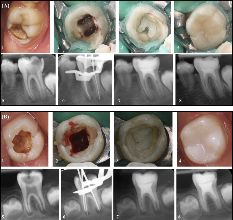
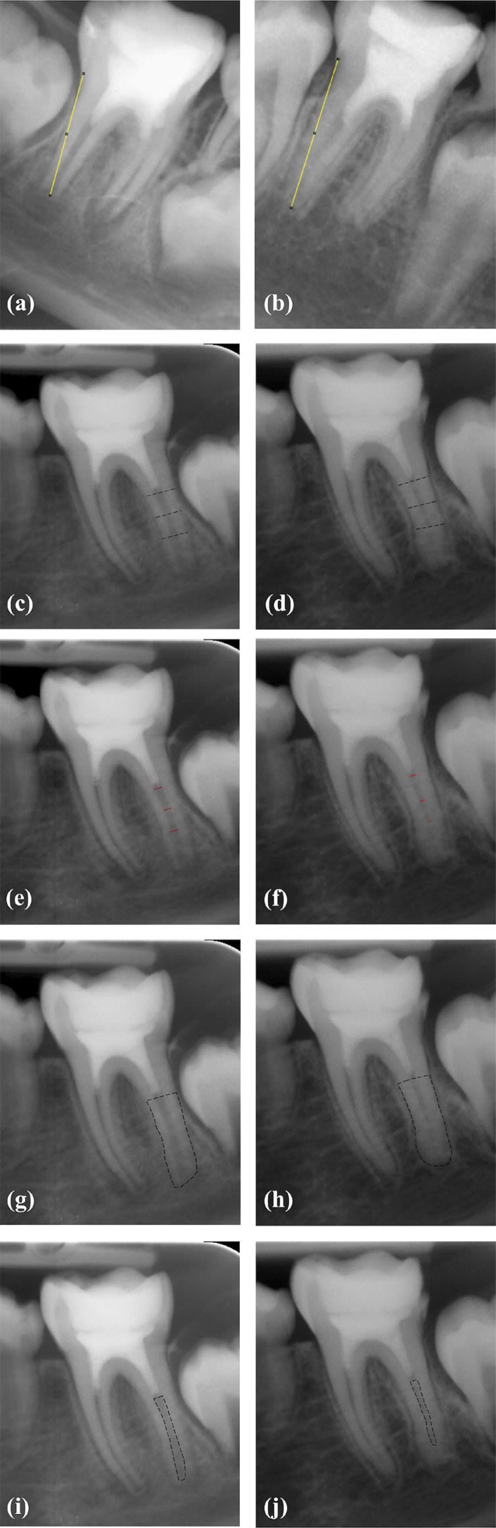
Methodology: The study included 20 patients with immature mandibular first molars exhibiting SIP/SAP. Participants were randomly assigned into two groups based on the applied intracanal medicament. Ca(OH)2 (n=10) was prepared by mixing it with sterile distilled water, while the same amount of powdered metronidazole and ciprofloxacin were mixed and combined with sterile distilled water for DAP (n=10). MMP-8 in periapical samples were measured at baseline and on the 14th day using immunofluorometric assay. Image-J software with TurboReg plug-in was utilized to determine changes in root length, root width, radiographic root area (RRA) during the 12-month follow-up period. Data were analyzed by SPSS 25.0 (p<.05).
Results: Significant increase in MMP-8 on the 14th day compared to baseline in both groups (p<0.001). There was no significant difference between the two groups in terms of the increase in MMP-8 (p>0.05). Root length significantly increased in both groups (p=0.001), with Ca(OH)2 showing a greater increase (p=0.046). Root width and RRA increased similarly in both groups at 12th month.
Conclusion: Both Ca(OH)2 and DAP applications resulted in a significant increase in periapical MMP-8 levels. Increase in radiographic root width and root area was similar between two groups, but Ca(OH)2 led to a significantly greater increase in root length. Further studies with larger sample sizes are necessary to validate our findings during REP of vital immature permanent mandibular molars. Clinical Trials database: NCT05581706.
Conflict of interest statement
Figures

References
-
- Berman LH, Hargreaves KM. Cohen's pathways of the pulp. Chicago: Elsevier Health Sciences; 2020. pp. 28–30.
Publication types
MeSH terms
Substances
Associated data
LinkOut - more resources
Full Text Sources
Medical
Miscellaneous
