. 2024 Sep 20:32:e20240168.
doi: 10.1590/1678-7757-2024-0168. eCollection 2024.

Effects of Thai propolis mixed in mineral trioxide aggregate on matrix metalloproteinase-2 expression and activity in inflamed human dental pulp cells

Affiliations

Effects of Thai propolis mixed in mineral trioxide aggregate on matrix metalloproteinase-2 expression and activity in inflamed human dental pulp cells

Nutnicha Tiyapitsanupaisan et al. J Appl Oral Sci. .

Abstract

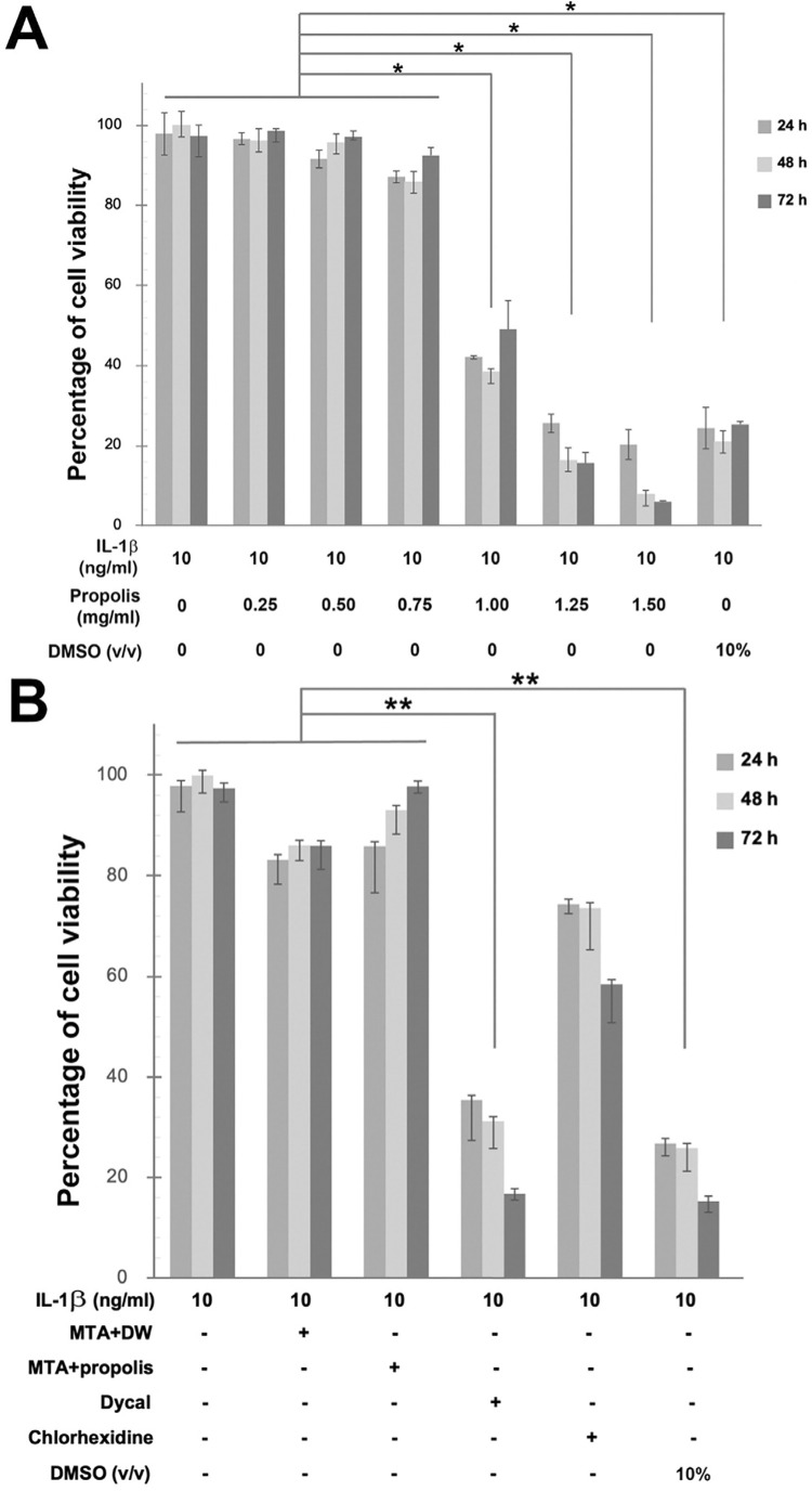
Objectives: This study sought to determine effects of Thai propolis extract mixed in mineral trioxide aggregate (MTA) on matrix metalloproteinase-2 (MMP-2) expression and its activity in inflamed human dental pulp cells (HDPCs).

Materials and methods: Interleukin-1β-primed HDPCs were treated with either the eluate of MTA mixed with distilled water, of MTA mixed with 0.75 mg/ml of the propolis extract, or of Dycal®, 0.75 mg/ml of the propolis extract, or 0.2% (v/v) of chlorhexidine for 24 or 72 h. The viability of HDPCs was determined by the PrestoBlue® cytotoxic assay. HDPCs' lysates were analyzed for MMP-2 mRNA expression by RT-qPCR, while their supernatants were measured for MMP-2 activity by gelatin zymography.

Results: At 24 and 72 h, a non-toxic dose of the propolis extract at 0.75 mg/ml by itself or mixed in MTA tended to reduce MMP-2 expression upregulated by MTA, while it further decreased the MMP-2 activity as compared to that of MTA mixed with distilled water. The MMP-2 activity of interleukin-1β-primed HDPCs treated with the eluate of the propolis extract mixed in MTA was significantly lower than that of interleukin-1β-primed HDPCs at 24 h (p=0.012). As a control, treatment with chlorhexidine significantly inhibited MMP-2 expression induced by MTA and MMP-2 activity enhanced by interleukin-1β (p<0.05). Treatment with Dycal® caused a significant increase in HDPC's death, resulting in a significant decrease in MMP-2 expression and activity (p<0.05).

Conclusions: MTA mixed with Thai propolis extract can reduce MMP-2 mRNA expression and activity when compared to MTA mixed with distilled water in inflamed HDPCs.


Conflict of interest statement

Conflict of interest: The authors declare no conflict of interest.

Figures

None
Graphical abstract
Figure 1
Figure 1. In vitro experimental designs of this study. Three flow charts (A, B, C) illustrating each research design of this study. Each experiment was repeated three times for each of the three different human dental pulp cells (HDPCs), isolated from three different donors (n = 3). D) The picture shows a vinylpolysiloxane-added silicone mold and the prepared MTA mixed with Thai propolis extract at 0.75 mg/ml which was immersed in DMEM for 24 h.
Figure 2
Figure 2. HDPCs viability test by PrestoBlue assay. Bar graphs demonstrating the percentages of cell viability upon treatment of IL-1β-primed HDPCs for 24, 48, or 72 h with indicated doses of Thai propolis extract (A) or with 100 ml of the eluate released from MTA mixed with distilled water (DW), that from MTA mixed with Thai propolis extract at 0.75 mg/ml, or that from Dycal®, or 100 ml of 0.2% (v/v) of chlorhexidine (B). Treatment for 24, 48, or 72 h with 10% (v/v) of dimethyl sulfoxide (DMSO) in A and B was used as positive control. Thai propolis extract at 0.75 mg/ml was demonstrated to be the highest, most non-toxic dose used for the subsequent experiments. Error bars = standard deviation; ** = p<0.01; * = p<0.05.
Figure 3
Figure 3. Microscopic imaging of HDPCs. Representative images showing cultured HDPCs upon treatment for 24 h (A, B, C) or 72 h (D, E, F) with 1.5 ml of the eluate released from MTA mixed with distilled water (A, D), from MTA mixed with Thai propolis extract at 0.75 mg/ml (B, E), or from Dycal® (C, F). White arrows in C, D, and F indicate rounded-shape dead cells in the culture.
Figure 4
Figure 4. A potential reduction of MMP-2 transcripts in MTA-treated HDPCs by propolis. A bar graph demonstrating folds of MMP-2 mRNA expression, normalized by GAPDH mRNA expression, in IL-1β-primed HDPCs treated for 24 h (light gray) or 72 h (dark gray) with 1.5 ml of the eluate released from MTA mixed with distilled water (DW), from MTA mixed with Thai propolis extract at 0.75 mg/ml, or from Dycal®, Thai propolis extract at 0.75 mg/ml, or 0.2% (v/v) of chlorhexidine relative to normalized MMP-2 mRNA expression of HDPCs that were neither stimulated nor treated, set to 1.0. Error bars = standard deviation; * = p<0.05 for comparisons at 24 h; ** = p<0.001 for comparisons at both 24 and 72 h. A = p<0.05, compared to unstimulated HDPCs at 24 h; B = p<0.05, compared to IL-1β-stimulated HDPCs that were not treated for 24 h; b = p<0.05, compared to IL-1β-stimulated HDPCs that were not treated for 72 h.
Figure 5
Figure 5. Gelatin Zymography for MMP-2 detection. Representative gelatin zymograms from the conditioned media of unstimulated HDPCS and that of IL-1β-primed HDPCs treated for 24 h (A) or 72 h (B) with 1.5 ml of the eluate released from MTA mixed with distilled water (DW), from MTA mixed with Thai propolis extract at 0.75 mg/ml, or from Dycal®, Thai propolis extract at 0.75 mg/ml, or 0.2% (v/v) of chlorhexidine. MTA mixed with either distilled water or Thai propolis extract dampened the degree of MMP-2 expression. Bar graphs below the zymograms illustrate changes in the percentages of MMP-2 activity, as measured by the densitometry of combined clear zones between pro-MMP-2 and active MMP-2, in the samples relative to that in the unstimulated HDPCs. Error bars = standard deviation; * = p<0.05. A = p<0.05, compared to unstimulated HDPCs at 24 h; B = p < 0.05, compared to IL-1β-stimulated HDPCs that were not treated for 24 h; a = p < 0.05, compared to unstimulated HDPCs at 72 h; b = p<0.05, compared to IL-1β-stimulated HDPCs that were not treated for 72 h, ns = non-significant, compared to IL-1β-stimulated HDPCs that were not treated for 72 h.
Figure 6
Figure 6. Significantly reduced levels of COX-2 and PGE2 when Thai propolis extract was used as vehicle for MTA preparation. Bar graphs illustrating folds of COX-2 mRNA expression, normalized by GAPDH mRNA expression, in the HDPCs’ lysates of samples in relation to that in the unstimulated HDPCs, set to 1.0 (A) and PGE2 levels in the HDPCs’ conditioned media in pg/ml (B). IL-1β-stimulated HDPCs were treated for 24 h (light gray) or 72 h (dark gray) with 1.5 ml of the eluate released from MTA mixed with distilled water (DW), from MTA mixed with Thai propolis extract at 0.75 mg/ml, or from Dycal®, or Thai propolis extract at 0.75 mg/ml. Error bars = standard deviation; * = p<0.05. A = p<0.05, compared to unstimulated HDPCs at 24 h; a = p<0.05, compared to unstimulated HDPCs at 72 h; B = p<0.05, compared to IL-1β-stimulated HDPCs that were not treated for 24 h; b = p<0.05, compared to IL-1β-stimulated HDPCs that were not treated for 72 h.

Similar articles

References

    1. Palosaari H, Pennington CJ, Larmas M, Edwards DR, Tjäderhane L, Salo T. Expression profile of matrix metalloproteinases (MMPs) and tissue inhibitors of MMPs in mature human odontoblasts and pulp tissue. Eur J Oral Sci. 2003;111(2):117–127. doi: 10.1034/j.1600-0722.2003.00026.x. - DOI - PubMed
    1. Charadram N, Farahani RM, Harty D, Rathsam C, Swain MV, Hunter N. Regulation of reactionary dentin formation by odontoblasts in response to polymicrobial invasion of dentin matrix. Bone. 2012;50(1):265–275. doi: 10.1016/j.bone.2011.10.03. - DOI - PMC - PubMed
    1. Tjäderhane L, Buzalaf MA, Carrilho M, Chaussain C. Matrix metalloproteinases and other matrix proteinases in relation to cariology: the era of 'dentin degradomics'. Caries Res. 2015;49(3):193–208. doi: 10.1159/000363582. - DOI - PubMed
    1. Mazzoni A, Mannello F, Tay FR, Tonti GA, Papa S, Mazzotti G, et al. Zymographic analysis and characterization of MMP-2 and -9 forms in human sound dentin. J Dent Res. 2007;86(5):436–440. doi: 10.1177/154405910708600509. - DOI - PubMed
    1. Veerayutthwilai O, Byers MR, Pham TT, Darveau RP, Dale BA. Differential regulation of immune responses by odontoblasts. Oral Microbiol Immunol. 2007;22(1):5–13. doi: 10.1111/j.1399-302X.2007.00310.x. - DOI - PubMed

Publication types

MeSH terms