. 2024 Dec 11;47(12):zsae225.
doi: 10.1093/sleep/zsae225.

Sleep state-dependent development of resting-state functional connectivity during the preterm period

Affiliations

Sleep state-dependent development of resting-state functional connectivity during the preterm period

Anna Shiraki et al. Sleep. .

Abstract

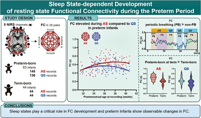
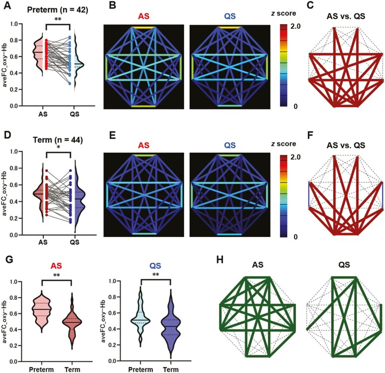
Study objectives: The brains of preterm infants exhibit altered functional connectivity (FC) networks, but the potential variation in sleep states and the impact of breathing patterns on FC networks are unclear. This study explores the evolution of resting-state FC from preterm to term, focusing on breathing patterns and distinguishing between active sleep (AS) and quiet sleep (QS).

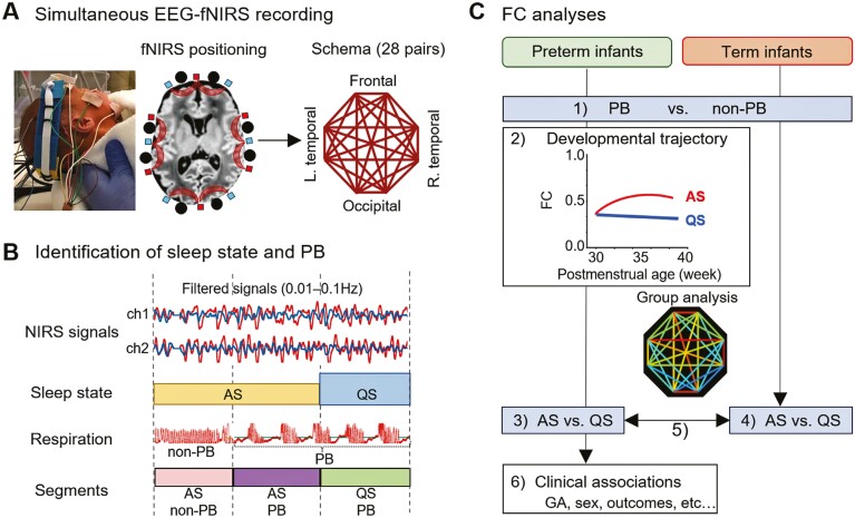
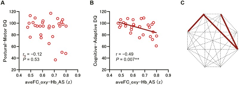
Methods: We recruited 63 preterm infants and 44 healthy-term infants and performed simultaneous electroencephalography and functional near-infrared spectroscopy. FC was calculated using oxy- and deoxyhemoglobin signals across eight channels. First, FC was compared between periodic breathing (PB) and non-PB segments. Then sleep state-dependent FC development was explored. FC was compared between AS and QS segments and between preterm infants at term and term-born infants in each sleep state. Finally, associations between FC at term, clinical characteristics, and neurodevelopmental outcomes in late infancy were assessed in preterm infants.

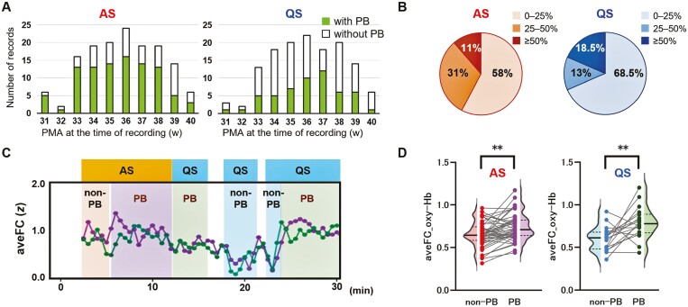
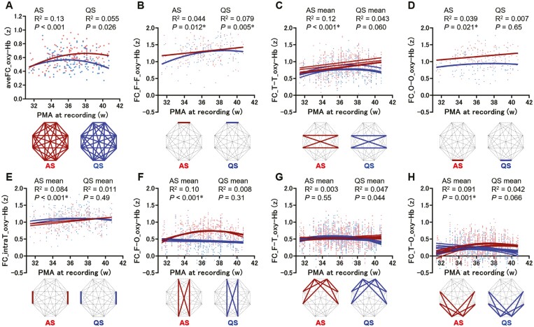
Results: In total, 148 records from preterm infants and 44 from term-born infants were analyzed. PB inflated FC values. After excluding PB segments, FC was found to be elevated during AS compared to QS, particularly in connections involving occipital regions. Preterm infants had significantly higher FC in both sleep states compared to term-born infants. Furthermore, stronger FC in specific connections during AS at term was associated with unfavorable neurodevelopment in preterm infants.

Conclusions: Sleep states play a critical role in FC development and preterm infants show observable changes in FC.

Keywords: electroencephalography; functional connectivity; near-infrared spectroscopy; outcome; periodic breathing; preterm infant; sleep state.


Figures

Graphical Abstract
Graphical Abstract
Figure 1.
Figure 1.
Overview of the study. (A) Simultaneous electroencephalography (EEG) and functional near-infrared spectroscopy (fNIRS) recording were performed. An eight-channel NIRS device was positioned around the infant’s head, as illustrated in MRI data acquired at 34 weeks postmenstrual age. The source and the detector locations are indicated by distinct colored squares, with each color representing a different type. Circles on shaded curves represent NIRS channels. Functional connectivity (FC) in each connection pair is represented as the schema. (B) FC was calculated based on filtered oxyhemoglobin and deoxy-hemoglobin data. Active sleep (AS) and quiet sleep (QS) states were determined every 30 seconds using EEG, electrooculogram, and respiratory data. Periodic breathing (PB) and non-PB segments were evaluated separately. Four groups were distinguished: AS non-PB, AS PB, QS non-PB, and QS PB. (C) The analyses performed were as follows: comparison of FC between non-PB and PB segments in AS and QS (with only non-PB segments used in subsequent analyses); analysis of the FC trajectory in preterm infants in both AS and QS; comparison of FC between AS and QS in preterm infants at term; comparison of FC between AS and QS in term-born infants; comparison of FC between preterm infants at term and term-born infants in both AS and QS; and exploration of the associations between FC at term and clinical characteristics, such as gestational age (GA), sex, and developmental outcomes, in preterm infants.
Figure 2.
Figure 2.
Periodic breathing (PB) in preterm infants. (A) Bar charts illustrating the numbers of records with and without PB according to postmenstrual age (PMA) at the time of recording in active sleep (AS, left) and quiet sleep (QS, right). (B) The proportion of time containing PB to the total recording time is depicted in pie charts for the records containing PB. (C) Representative 30-minute time-course of average functional connectivity (aveFC). Oxy-hemoglobin (Hb) data and deoxy-Hb data are represented by different colored dots, both showing similar trends. (D) AveFC for oxy-Hb signals in non-PB and PB segments in AS (left) and QS (right). Data from 49 AS and 18 QS records, including both non-PB and PB segments, were compared using a paired t-test (AS, p < .001; QS, p = .004). **p < .01.
Figure 3.
Figure 3.
Developmental trajectories of functional connectivity in preterm infants (oxy-hemoglobin [Hb]). (A–H) Scatter plots and regression lines show the average functional connectivity (aveFC) of 28 connections (A) and the functional connectivity (FC) for individual connections (B–H) in oxy-Hb signals according to sleep state. FC values were calculated using non-periodic breathing data extracted from 143 records obtained during active sleep (AS) and 132 records obtained during quiet sleep (QS). The x-axis indicates postmenstrual age (PMA) at the time of recording, and the y-axis indicates aveFC (A) and FC (B–H) in oxy-Hb signals (z scores). The FC in AS and QS are represented in different colors. The connections were categorized into seven types (B–H), as shown in the lower column, based on brain regions and FC values. Linear or nonlinear regression analysis was conducted depending on the mean FC for each connection type. P-values calculated before the false discovery rate (FDR) adjustment are shown. *p < .05 after FDR adjustment.
Figure 4.
Figure 4.
Disparities in functional connectivity (FC) between active sleep (AS) and quiet sleep (QS), and between preterm infants at term and term-born infants. (A) The average functional connectivity (aveFC) in oxy-hemoglobin (Hb) signals was compared between AS and QS in preterm infants at term using a paired t-test (p < .001). (B) Analysis of FC using oxy-Hb data from 42 preterm infants at term, in AS and QS, with FC strength indicated by the color bar. (C) Comparison of FC between AS and QS in preterm infants at term. A paired t-test was conducted to analyze the FC of all connections and connections that were significantly stronger in AS are depicted by bold solid lines. (D) AveFC for oxy-Hb signals in term-born infants: comparison between AS and QS using a paired t-test (p = .016). (E) Analysis of FC using oxy-Hb data from 44 term-born infants in AS and QS. (F) Comparison of FC between AS and QS in term-born infants. A paired t-test was conducted to analyze all FC connections; connections that were significantly stronger in AS are depicted by bold solid lines, and connections that were significantly weaker in AS are represented by thin solid lines (bilateral intra-hemispheric temporal connections). (G) AveFC for oxy-Hb signals recorded during AS and QS were compared between preterm infants at term and term-born infants using a two-sample t-test (AS, p < .001; QS, p < .001). (H) Comparison of FC between preterm infants at term and term-born infants in AS and QS. A two-sample t-test was conducted to analyze all FC connections and connections that were significantly stronger in preterm infants than in term-born infants are depicted by solid lines. No FC connections were significantly weaker in preterm infants than in term-born infants. Two-tailed p-values < .05 for aveFC, and for FC in individual connections after false discovery rate adjustment, were considered statistically significant. *p < .05; **p < .01. Detailed FC data are presented in Supplementary Tables S1–S4.
Figure 5.
Figure 5.
Association between the average functional connectivity (aveFC) at term and developmental quotient (DQ) in preterm infants. (A) Correlation between the aveFC for oxy-hemoglobin (Hb) signals at term in active sleep (AS) and the postural–motor DQ at 10 months of corrected age in preterm infants. (B) Correlation between the aveFC for oxy-Hb at term in AS and the cognitive–adaptive DQ at 10 months of corrected age in preterm infants. (C) Connections with functional connectivity (FC) values that were significantly negatively correlated (two-tailed p-value < .05 after false discovery rate adjustment) with the cognitive–adaptive DQ at 10 months of corrected age are represented by solid lines. Note that no FC connections showed a positive correlation with the cognitive–adaptive DQ. **p < .01.

Similar articles

Cited by

References

    1. Luhmann HJ, Kanold PO, Molnár Z, Vanhatalo S.. Early brain activity: translations between bedside and laboratory. Prog Neurobiol. 2022;213:102268. doi: https://doi.org/10.1016/j.pneurobio.2022.102268 - DOI - PMC - PubMed
    1. Molnar Z, Luhmann HJ, Kanold PO.. Transient cortical circuits match spontaneous and sensory-driven activity during development. Science. 2020;370(6514):eabb2153. doi: https://doi.org/10.1126/science.abb2153 - DOI - PMC - PubMed
    1. Inder TE, Volpe JJ, Anderson PJ.. Defining the neurologic consequences of preterm birth. N Engl J Med. 2023;389(5):441–453. doi: https://doi.org/10.1056/NEJMra2303347 - DOI - PubMed
    1. Pierrat V, Marchand-Martin L, Arnaud C, et al.; EPIPAGE-2 writing group. Neurodevelopmental outcome at 2 years for preterm children born at 22 to 34 weeks’ gestation in France in 2011: EPIPAGE-2 cohort study. BMJ. 2017;358:j3448. doi: https://doi.org/10.1136/bmj.j3448 - DOI - PMC - PubMed
    1. Cheong JLY, Olsen JE, Lee KJ, et al.; Victorian Infant Collaborative Study Group. Temporal trends in neurodevelopmental outcomes to 2 years after extremely preterm birth. JAMA Pediatr. 2021;175(10):1035–1042. doi: https://doi.org/10.1001/jamapediatrics.2021.2052 - DOI - PMC - PubMed

Publication types