
Virulence of Burkholderia pseudomallei ATS2021 Unintentionally Imported to United States in Aromatherapy Spray

Christopher K Cote et al. Emerg Infect Dis. 2024 Oct.

Abstract

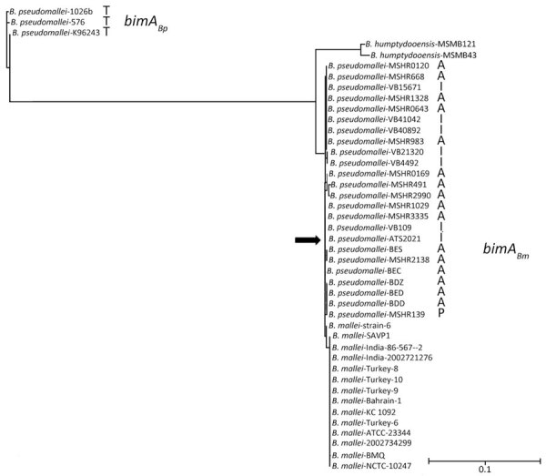
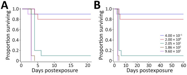
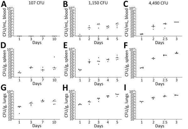
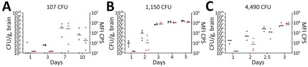
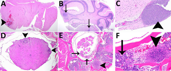
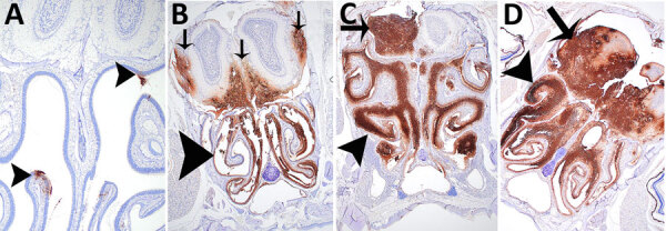
In the United States in 2021, an outbreak of 4 cases of Burkholderia pseudomallei, the etiologic agent of melioidosis and a Tier One Select Agent (potential for deliberate misuse and subsequent harm), resulted in 2 deaths. The causative strain, B. pseudomallei ATS2021, was unintentionally imported into the United States in an aromatherapy spray manufactured in India. We established that ATS2021 represents a virulent strain of B. pseudomallei capable of robust formation of biofilm at physiologic temperatures that may contribute to virulence. By using mouse melioidosis models, we determined median lethal dose estimates and analyzed the bacteriologic and histopathologic characteristics of the organism, particularly the potential neurologic pathogenesis that is probably associated with the bimABm allele identified in B. pseudomallei strain ATS2021. Our data, combined with previous case reports and the identification of endemic B. pseudomallei strains in Mississippi, support the concept that melioidosis is emerging in the United States.

Keywords: ATS2021; Burkholderia pseudomallei; Indian strain; United States; bacteria; biofilm; imported; melioidosis; mice; neurologic.


Figures

Figure 1
Figure 1
Phylogenetic tree based on bimABp and bimABm alleles from strains of Burkholderia pseudomallei, B. mallei, and B. humptydooensis, showing location of B. mallei ATS2021 (arrow), the causative strain in an outbreak of 4 cases, 2 of them fatal, in the United States in 2021. NGPhylogeny.fr (12) was used to build the tree in “A la Carte” mode, and it used MUSCLE for multiple alignment, Gblocks for automatic alignment curation (https://NGphylogeny.fr for both), PhyML-SMS (https://www.atgc-montpellier.fr) for tree inference, and exported to the Interactive Tree Of Life (iTOL; https://itol.embl.de) for display and manipulation. B. pseudomallei strains were isolated in Thailand (T), Australia (A), India (I), and Papua New Guinea (P). Scale bar indicates number of substitutions per site. bimA, Burkholderia intracellular motility factor A.
Figure 2
Figure 2
Biofilm formation of Burkholderia pseudomallei strains ATS2021, the causative strain in an outbreak of 4 cases, 2 of them fatal, in the United States in 2021, and K96243 under multiple test conditions. Biofilm formation of both strains was assessed by crystal violet staining as measured by OD600. A) One day at room temperature; B) 1 day at 37°C; C) 2 days at room temperature; D) 2 days at 37°C. Error bars represent the SEs from mean values determined from 3 separate assays. p values determined by linear mixed effects model. M9G, M9 + glucose; M9GC, 0.2% glucose to M9 and casamino acids; OD600, optical density at 600 nm.
Figure 3
Figure 3
Biofilm production for Burkholderia pseudomallei strain ATS2021, the causative strain in an outbreak of 4 cases, 2 of them fatal, in the United States in 2021, in relation to other previously characterized B. pseudomallei clinical isolates. Biofilm formation of bacterial strains was assessed by crystal violet staining as measured by OD600. Biofilm was allowed to form after static growth in LB+4% glycerol for 1 day at 37°C. Clinical isolates used in this assay originated from Thailand (red bars) or Australia (green bars). Error bars represent the SEs from mean values determined from 4 independent assays. ATS2021 formed significantly more biofilm under these conditions compared to all isolates except MSHR5848 as determined by a linear mixed effects model. OD600, optical density at 600 nm.
Figure 4
Figure 4
Kaplan-Meier survival plots calculated for C57BL/6 mice exposed to aerosolized Burkholderia pseudomallei ATS2021, the causative strain in an outbreak of 4 cases, 2 of them fatal, in the United States in 2021. A) Survival after 21 days; B) survival after 60 days. N = 10 per aerosolized dose of B. pseudomallei. The day 21 survival rates from highest to lowest calculated inhaled dose of B. pseudomallei ATS2021 (4,490 CFU, 1,150 CFU, 107 CFU, 13 CFU, 1 CFU) as depicted in panel A are 0/10, 0/10, 4/10, 8/10, and 10/10. The day 60 survival rates from highest to lowest calculated inhaled dose of B. pseudomallei ATS2021 as depicted in panel B are 0/10, 0/10, 0/10, 3/10, and 9/10.
Figure 5
Figure 5
Kaplan-Meier survival plots calculated for BALB/c mice exposed to aerosolized Burkholderia pseudomallei ATS2021, the causative strain in an outbreak of 4 cases, 2 of them fatal, in the United States in 2021. A) Survival after 21 days; B) survival after 60 days. N = 10 per aerosolized dose of B. pseudomallei. The day 21 survival rates from highest to lowest calculated inhaled dose of B. pseudomallei ATS2021 (960 CFU, 186 CFU, 21 CFU, 2 CFU, 0.4 CFU) as depicted in panel A are 0/10, 0/10, 1/10, 8/10, and 9/10. The day 60 survival rates as depicted in panel B are 0/10, 0/10, 1/10, 8/10, and 9/10.
Figure 6
Figure 6
Serial sampling experiment to investigate bacterial dissemination in blood, spleens, and lungs of C57BL/6 mice at different challenge doses of aerosolized Burkholderia pseudomallei strain ATS2021, the causative strain in an outbreak of 4 cases, 2 of them fatal, in the United States in 2021. C57BL/6 mice were estimated to have inhaled various doses on day 0. A subset of mice was then deeply anesthetized for a terminal blood collection, euthanized, and then the spleens and lungs were removed to determine the bacteriologic burden in each organ at each time point indicated. Bacterial counts are shown for whole blood (A–C), spleen homogenate (D–F), and lung homogenate (G–I). N = 4 for each time point. The CFU burden is shown for each mouse; the geometric mean is depicted with the horizontal bar. Limit of detection is ≈100 CFU/mL of blood and 5 CFU/organ.
Figure 7
Figure 7
Serial sampling experiment to investigate bacterial dissemination in brains of C57BL/6 mice at different challenge doses of aerosolized Burkholderia pseudomallei strain ATS2021, the causative strain in an outbreak of 4 cases, 2 of them fatal, in the United States in 2021. C57BL/6 mice were estimated to have inhaled various doses of aerosolized Burkholderia pseudomallei ATS2021 on day 0: A) 107 CFU, B) 1,150 CFU, and C) 4,490 CFU. A subset of mice was then deeply anesthetized for a terminal blood collection, euthanized, and then brains were removed to determine the bacteriologic burden in each brain at each time point indicated. N = 4 for each time point. The CFU burden is shown for each mouse, depicted as a black circle; geometric mean is depicted with the horizontal bar. The brain homogenates were exposed to ≈21 kGy of gamma radiation, proven sterile, and then subjected to the B. pseudomallei capsule-specific immunodiagnostic assay. The MFI of each mouse is depicted by a red circle, and the geometric mean is depicted with the horizontal bar. Limit of detection is ≈5 CFU/organ. The detection of capsule in the irradiated brain homogenate is depicted in red and is displayed as MFI. MFI, mean fluorescent intensity.
Figure 8
Figure 8
Hematoxylin and eosin staining of head sections of C57BL/6 mice exposed to aerosolized Burkholderia pseudomallei strain ATS2021, the causative strain in an outbreak of 4 cases, 2 of them fatal, in the United States in 2021. Shown are the nasal cavity (including nasal turbinates, nasal septum, respiratory and olfactory epithelium, lamina propria with supporting tissues and glands, nerve bundles, and nasal air passages); cribriform plate (bone and olfactory/trigeminal nerves); and cranial vault with olfactory bulb. A) Day 1 after exposure, dose 107 CFU, showing nasal turbinates (arrowhead), cribriform plate (asterisks), and olfactory bulb (arrow) that are essentially normal. Original magnification ×2. B) Day 2 after exposure, dose 4,490 CFU, showing mild necrosuppurative rhinitis (arrowheads) with edema, cellular debris and suppurative inflammation in few nasal air passages (arrows). Original magnification ×2. C) Day 3 after exposure, dose 4,490 CFU, showing multifocal moderate necrosuppurative rhinitis (arrowheads) and necrotizing and hemorrhagic meningoencephalitis of the olfactory bulb (arrows). Original magnification 2. D) Day 4 after exposure, dose 1,150 CFU. There is diffuse necrosuppurative rhinitis of the nasal turbinates (arrowheads), showing extensive necrosis and hemorrhage of the olfactory bulb with a small portion of recognizable neural tissue evident (arrow). Original magnification ×2.
Figure 9
Figure 9
Histopathologic analyses of the neurologic system of C57BL/6 mice after inhalation of Burkholderia pseudomallei strain ATS2021, the causative strain in an outbreak of 4 cases, 2 of them fatal, in the United States in 2021. A) Day 5 after exposure, dose 1,150 CFU. Cerebrum showing focally extensive necrotizing and hemorrhagic meningoencephalitis (arrowhead). Hematoxylin and eosin (HE) stain; original magnification ×2. B) Day 9 after exposure, dose 107 CFU. Pons and cerebellum. There is multifocal necrotizing meningoencephalitis (arrows). HE stain; original magnification ×2. C) Day 10 after exposure, dose 1,150 CFU. Cerebrum with olfactory peduncle filled with viable and degenerate neutrophils with no recognizable peduncular tissue (arrowhead). HE stain; original magnification ×20. D) Day 9 after exposure, 107 CFU. Spinal cord, thoracic, shows multifocal meningomyelitis (arrowheads). HE stain; original magnification ×4. E) Day 5 after exposure, 1,150 CFU. Spinal cord and vertebrae, lumbar at cauda equina, show is multifocal suppurative perineuritis of spinal nerves (arrows). Note the necrotizing lesion within the vertebral bone marrow (arrowhead) HE stain; original magnification ×10. F) Day 4 after exposure, dose 1,150 CFU. Spinal cord and vertebra, thoracic, show necrotizing osteomyelitis (arrowhead). Note the loss of distinction of bone marrow cells compared to normal cells of the bone marrow (arrow). HE stain; original magnification ×40.
Figure 10
Figure 10
Immunohistochemical analyses of head sections of C57BL/6 mice exposed to aerosolized Burkholderia pseudomallei strain ATS2021, the causative strain in an outbreak of 4 cases, 2 of them fatal, in the United States in 2021. Shown are the nasal cavity (including nasal turbinates, nasal septum, respiratory and olfactory epithelium, lamina propria with supporting tissues and glands, nerve bundles, and nasal air passages); cribriform plate (bone and olfactory/trigeminal nerves); and cranial vault with olfactory bulb. A) Day 1 after exposure, dose 1,150 CFU, shows multifocal minimal positivity of the nasal cavity epithelium (arrowheads), which is most likely olfactory epithelium of the ethmoid turbinate. Original magnification ×2. B) Day 3 after exposure, dose 4,490 CFU, showing diffuse marked positivity of the nasal cavity epithelium (arrowhead) and multifocal moderate positivity of the olfactory bulb (arrows). Original magnification ×2. C) Day 6 after exposure, dose 107 CFU, showing diffuse severe positivity of the nasal cavity epithelium (arrowhead) and multifocal marked positivity of the olfactory bulb (arrow). Original magnification ×2. D) Day 4 after exposure, 1,150 CFU, showing diffuse severe positivity of the nasal cavity (arrowhead) and olfactory bulb (arrow). Originalmagnification ×2.
Figure 11
Figure 11
Immunohistochemical analyses of the neurologic system of C57BL/6 mice exposed to aerosolized Burkholderia pseudomallei strain ATS2021, the causative strain in an outbreak of 4 cases, 2 of them fatal, in the United States in 2021. A) Day 6 after exposure, dose 107 CFU. Cerebrum shows multifocal moderate positivity (arrows). Original magnification ×2. B) Day 6 after exposure, dose 107 CFU. Cerebellum shows multifocal moderate positivity (arrow). Original magnification ×10. C) Day 5 after exposure, dose 1,150 CFU. Lumbar spinal cord at cauda equina and vertebra shows multifocal mild perineural positivity (arrows) of spinal nerves at cauda equina and marked vertebral bone marrow positivity (arrowheads). Original magnification ×10. D) Day 9 after exposure, dose 107 CFU. Spinal cord, cervical, shows focal moderate positivity (arrowheads). Original magnification ×20.
Figure 12
Figure 12
Profiling of differentially expressed genes revealing downregulation of markers associated with oligodendrocytes after exposure to aerosolized Burkholderia pseudomallei strain ATS2021, the causative strain in an outbreak of 4 cases, 2 of them fatal, in the United States in 2021. A) Significant downregulation or decreased expression of genes involved in oligodendrocyte function was observed in select challenge groups of mice on days 1,3, and 5 after challenge. Values are normalized to unchallenged control mice and represent 4 mice per group. Significance based on a 2-way analysis of variance. Error bars indicate 95% CIs. B) Heat map of differentially expressed genes associated with NanoString oligodendrocyte cell profiler panel (https://nanostring.com). Fold change is linear, and dots indicate gene changes that were above the significance threshold. C) Expression changes over time in select genes associated with oligodendrocyte function for the 1,150 CFU challenge group. In panels A and C, error bars represent standard deviation.

Similar articles

Cited by

References

    1. Currie BJ. Melioidosis: evolving concepts in epidemiology, pathogenesis, and treatment. Semin Respir Crit Care Med. 2015;36:111–25. 10.1055/s-0034-1398389 - DOI - PubMed
    1. Currie BJ, Meumann EM, Kaestli M. The expanding global footprint of Burkholderia pseudomallei and melioidosis. Am J Trop Med Hyg. 2023;108:1081–3. 10.4269/ajtmh.23-0223 - DOI - PMC - PubMed
    1. Chantratita N, Phunpang R, Yarasai A, Dulsuk A, Yimthin T, Onofrey LA, et al. Characteristics and one year outcomes of melioidosis patients in northeastern Thailand: a prospective, multicenter cohort study. Lancet Reg Health Southeast Asia. 2023;9:9. 10.1016/j.lansea.2022.100118 - DOI - PMC - PubMed
    1. Currie BJ, Mayo M, Ward LM, Kaestli M, Meumann EM, Webb JR, et al. The Darwin Prospective Melioidosis Study: a 30-year prospective, observational investigation. Lancet Infect Dis. 2021;21:1737–46. 10.1016/S1473-3099(21)00022-0 - DOI - PubMed
    1. Currie BJ, Fisher DA, Howard DM, Burrow JN. Neurological melioidosis. Acta Trop. 2000;74:145–51. 10.1016/S0001-706X(99)00064-9 - DOI - PubMed

LinkOut - more resources