

Distribution characteristics of Purkinje fibres in the canine left ventricle
- PMID: 39320327
- PMCID: PMC11423647
- DOI: 10.1111/jcmm.70117
Distribution characteristics of Purkinje fibres in the canine left ventricle
Erratum in
-
Correction to "Distribution characteristics of Purkinje fibres in the canine left ventricle".J Cell Mol Med. 2024 Nov;28(21):e70164. doi: 10.1111/jcmm.70164. J Cell Mol Med. 2024. PMID: 39530357 Free PMC article. No abstract available.
Abstract
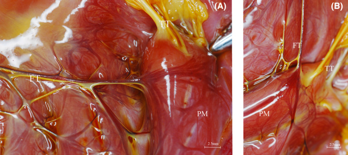
Purkinje-related ventricular arrhythmias have been increasingly reported, and with the development of catheter ablation techniques, intervention for Purkinje-related arrhythmias has been shown to be effective. The characteristics of Purkinje fibres orientation in the 12 canine left ventricles were observed at a gross level by staining the endocardium with Lugol's solution. Purkinje fibres were observed microscopically by HE, Masson's, PAS glycogen, and Cx40 immunohistochemical staining. Staining was successful, and the transverse orientation characteristics of Purkinje fibres were observed by Lugol's staining, and the longitudinal distribution was observed microscopically. The distribution of Purkinje fibres in the canine left ventricle is 'graded', 'layered', and 'networked', which can guide catheter ablation of Purkinje-related ventricular arrhythmia.
Keywords: anatomy; iodine staining; morphology; purkinje fibres.
© 2024 The Author(s). Journal of Cellular and Molecular Medicine published by Foundation for Cellular and Molecular Medicine and John Wiley & Sons Ltd.
Conflict of interest statement
The authors declare that they have no known competing financial interests or personal relationships that could have appeared to influence the work reported in this paper.
Figures


References
-
- Ishii K, Koga Y, Nakamura K, et al. Clinical application of a new vital staining method for the conduction system during heart operations. J Thorac Cardiovasc Surg. 1988;95(4):592‐597. - PubMed
MeSH terms
Substances
LinkOut - more resources
Full Text Sources
