

Clinical outcomes of percutaneous coronary intervention for severely calcified lesions: comparison between the morphologies of severely calcified coronary lesions
- PMID: 39320431
- PMCID: PMC11923015
- DOI: 10.1007/s00380-024-02466-7
Clinical outcomes of percutaneous coronary intervention for severely calcified lesions: comparison between the morphologies of severely calcified coronary lesions
Abstract
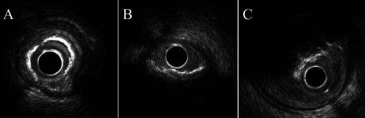
Existing studies evaluating the comparison of clinical outcome of percutaneous coronary intervention (PCI) for severe calcified coronary lesions are limited, and the clinical outcomes of PCI for different morphologies of calcified lesions are controversial. Overall, consecutive 576 lesions with severe calcification that were treated with PCI from 2010 to 2021 at Nagoya Heart Center were investigated. All lesions were assessed using invasive coronary angiogram (CAG) or computed tomography-CAG at 12 months after DES implantation. We divided the patients into three groups based on the results of intravascular ultrasound (IVUS) imaging (concentric calcified lesion [CC] n = 273, eccentric calcified lesion [EC] n = 217, calcified nodule [CN] n = 86). The clinical and angiographic outcomes of each group were investigated retrospectively to compare the prognosis between the three groups and identify predictive factors for the device-oriented composite end points (DoCE). There were no differences in patient characteristics among the three groups, except that there were significantly more patients on dialysis in the CN group. The incidence of DoCE was significantly higher in the CN group than in the other groups (CC; 18.3% vs. EC; 23.5% vs. CN; 36.0%; Log-Rank test; p = 0.001). Cox regression analysis showed that the independent predictors of DoCE were CN, insulin use, hemodialysis, right coronary artery lesions, and calcium cracks. The incidence of DoCE was significantly higher in the CN group. Calcium cracks are crucial for improving outcomes in severely calcified lesions, being key predictors of DoCE.
Keywords: Calcification; Drug-eluting stent; Percutaneous coronary intervention.
© 2024. The Author(s).
Conflict of interest statement
Declarations. Conflict of interest: All authors have no conflicts of interest to declare.
Figures


Comment in
-
Enhancing PCI strategies for severely calcified coronary lesions: gaps and new directions.Heart Vessels. 2025 May;40(5):456-457. doi: 10.1007/s00380-024-02494-3. Epub 2024 Nov 23. Heart Vessels. 2025. PMID: 39579205 No abstract available.
References
-
- Guedeney P, Claessen BE, Mehran R, Mintz GS, Liu M, Sorrentino S, Giustino G, Farhan S, Leon MB, Serruys PW, Smits PC, von Birgelen C, Ali ZA, Généreux P, Redfors B, Madhavan MV, Ben-Yehuda O, Stone GW (2020) Coronary calcification and long-term outcomes according to drug-eluting stent generation. JACC Cardiovasc Interv 13(12):1417–1428 - PubMed
-
- Kuramitsu S, Ohya M, Shinozaki T, Otake H, Horie K, Kawamoto H, Yamanaka F, Natsuaki M, Shiomi H, Nakazawa G, Ando K, Kadota K, Saito S, Kimura T (2019) Risk factors and long-term clinical outcomes of second-generation drug-eluting stent thrombosis. Circ Cardiovasc Interv 12(6):e007822 - PubMed
-
- Madhavan MV, Tarigopula M, Mintz GS, Maehara A, Stone GW, Généreux P (2014) Coronary artery calcification: pathogenesis and prognostic implications. J Am Coll Cardiol 63(17):1703–1714 - PubMed
-
- Fujino A, Mintz GS, Lee T, Hoshino M, Usui E, Kanaji Y, Murai T, Yonetsu T, Matsumura M, Ali ZA, Jeremias A, Moses JW, Shlofmitz RA, Kakuta T, Maehara A (2018) Predictors of calcium fracture derived from balloon angioplasty and its effect on stent expansion assessed by optical coherence tomography. JACC Cardiovasc Interv 11(10):1015–1017 - PubMed
-
- Kubo T, Shimamura K, Ino Y, Yamaguchi T, Matsuo Y, Shiono Y, Taruya A, Nishiguchi T, Shimokado A, Teraguchi I, Orii M, Yamano T, Tanimoto T, Kitabata H, Hirata K, Tanaka A, Akasaka T (2015) Superficial calcium fracture after PCI as assessed by OCT. JACC Cardiovasc Imaging 8(10):1228–1229 - PubMed
Publication types
MeSH terms
LinkOut - more resources
Full Text Sources
Medical
Miscellaneous
