

Complication rates after autologous cranioplasty following decompressive craniectomy
- PMID: 39320557
- PMCID: PMC11424706
- DOI: 10.1007/s00701-024-06282-w
Complication rates after autologous cranioplasty following decompressive craniectomy
Abstract
Objective: The reimplantation of autologous bone grafts after decompressive craniectomy (DC) is still up for debate. The objective of this study was to analyze the surgical revision rate for autologous cranioplasties in our center, aiming to identify predictors for procedure-related-complications.
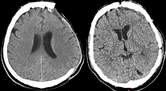
Methods: A retrospective single-center study was conducted for adult patients who underwent autologous cranioplasty after DC. The primary endpoint was the complication rate in terms of surgical revision and removal of the bone graft: infection, new onset seizures, dislocation, haemorrhage, osteolysis, wound dehiscence and cerebrospinal fluid (CSF) fistula. Demographic data, medical records, surgical reports and imaging studies were analysed and risk factors for complications were evaluated.
Results: 169 consecutive patients were included. The median interval between DC and cranioplasty was 84 days. Mean age was 51 ± 12.4 years. 26 patients (15.3%) had revision surgery for following reasons. n = 9 implant dislocations (5.3%), n = 7 osteolysis (3.6%), n = 6 infections (3.6%), n = 5 had re-bleedings (3%), n = 5 wound dehiscences (3%), and n = 2 CSF fistulas (1.2%). 18 patients developed new seizures (10.7%). Bi- and multivariate analysis revealed three independent risk factors, simultaneous ventriculo-peritoneal (VP) shunting increased the risk for material dislocation (p < 0.001); large bone grafts (> 193.5 cm2) increased the risk for osteolysis (p = 0.001) and bifrontal cranioplasties were associated with higher risk for infections (p = 0.04).
Conclusion: The complication rates in our study were comparable to previously reported data for autologous or artificial cranioplasties. As osteolysis was correlated to larger bone grafts, a synthetic alternative should be considered in selected cases.
Keywords: Autologous cranioplasty; Bone flap resorption; Complications; Cranial defect; Decompressive craniectomy; Infection; Risk factors; Skull reconstruction.
© 2024. The Author(s).
Conflict of interest statement
The authors have no competing interests to declare that are relevant to the content of this article.
Figures
Similar articles
-
The role of autologous bone in cranioplasty. A systematic review of complications and risk factors by using stored bone.Acta Neurochir (Wien). 2024 Nov 4;166(1):438. doi: 10.1007/s00701-024-06312-7. Acta Neurochir (Wien). 2024. PMID: 39495337
-
A systematic review and meta-analysis of factors involved in bone flap resorption after decompressive craniectomy.Neurosurg Rev. 2022 Jun;45(3):1915-1922. doi: 10.1007/s10143-022-01737-z. Epub 2022 Jan 21. Neurosurg Rev. 2022. PMID: 35061139
-
Complications and cosmetic outcomes of materials used in cranioplasty following decompressive craniectomy-a systematic review, pairwise meta-analysis, and network meta-analysis.Acta Neurochir (Wien). 2022 Dec;164(12):3075-3090. doi: 10.1007/s00701-022-05251-5. Epub 2022 May 20. Acta Neurochir (Wien). 2022. PMID: 35593924
-
Complications After In Vivo and Ex Vivo Autologous Bone Flap Storage for Cranioplasty: A Comparative Analysis of the Literature.World Neurosurg. 2016 Dec;96:510-515. doi: 10.1016/j.wneu.2016.09.025. Epub 2016 Sep 16. World Neurosurg. 2016. PMID: 27647038
-
Autologous Bone Is Inferior to Alloplastic Cranioplasties: Safety of Autograft and Allograft Materials for Cranioplasties, a Systematic Review.World Neurosurg. 2018 Sep;117:443-452.e8. doi: 10.1016/j.wneu.2018.05.193. Epub 2018 Jun 5. World Neurosurg. 2018. PMID: 29879511
Cited by
-
Rapid neuralized and vascularized osteogenesis in infected bone defect using biomimetic biomineralized and antibacterial hydrogels.Front Bioeng Biotechnol. 2025 May 21;13:1611639. doi: 10.3389/fbioe.2025.1611639. eCollection 2025. Front Bioeng Biotechnol. 2025. PMID: 40470505 Free PMC article.
References
-
- Alkhaibary A, Alharbi A, Abbas M et al (2020) Predictors of Surgical Site Infection in Autologous Cranioplasty: A Retrospective Analysis of Subcutaneously Preserved Bone Flaps in Abdominal Pockets. World Neurosurg 133:e627–e632. 10.1016/j.wneu.2019.09.120 - PubMed
-
- Charlson ME, Pompei P, Ales KL, MacKenzie CR (1987) A new method of classifying prognostic comorbidity in longitudinal studies: development and validation. J Chronic Dis 40(5):373–383. 10.1016/0021-9681(87)90171-8 - PubMed
-
- De Bonis P, Frassanito P, Mangiola A, Nucci CG, Anile C, Pompucci A (2012) Cranial repair: how complicated is filling a “hole”? J Neurotrauma 29(6):1071–1076. 10.1089/neu.2011.2116 - PubMed
Publication types
MeSH terms
LinkOut - more resources
Full Text Sources
Medical
Research Materials
