

Corneal densitometry measurements comparison between anterior segment OCT and scheimpflug imaging
- PMID: 39320570
- PMCID: PMC11424699
- DOI: 10.1007/s10792-024-03309-0
Corneal densitometry measurements comparison between anterior segment OCT and scheimpflug imaging
Abstract
Purpose: To evaluate and compare the repeatability of corneal densitometry (CD) measurements obtained using both an anterior-segment optical coherence tomography (AS-OCT) device and a Scheimpflug camera system, while also assessing the level of agreement. The study also sought to investigate the correlation of CD with age, gender, and central corneal thickness (CCT) in normal eyes.
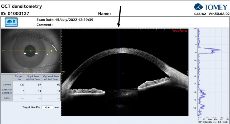
Methods: CD measurements were obtained using the Casia 2 and the Pentacam AXL Wave. Data were collected on Total Corneal Densitometry and 4 concentric corneal annular areas, these are referred to as zone 1, denoting the central area, through to zone 4, designating the outermost peripheral region. Repeatability was assessed using intra-session test-retest variability, coefficient of variation (CoV), and intraclass correlation coefficient (ICC). The agreement was evaluated using Bland-Altman plots. Correlation analysis was performed between CD, age, gender, and CCT.
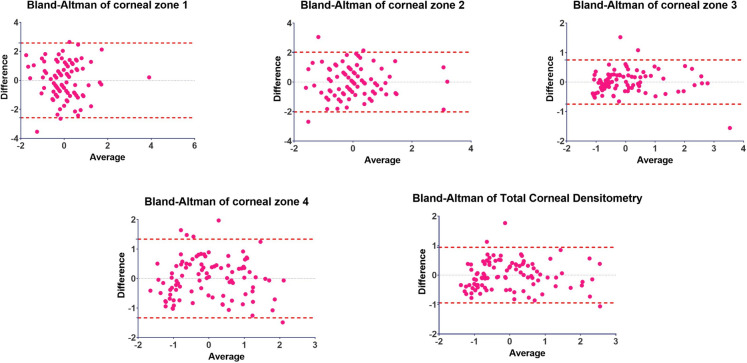
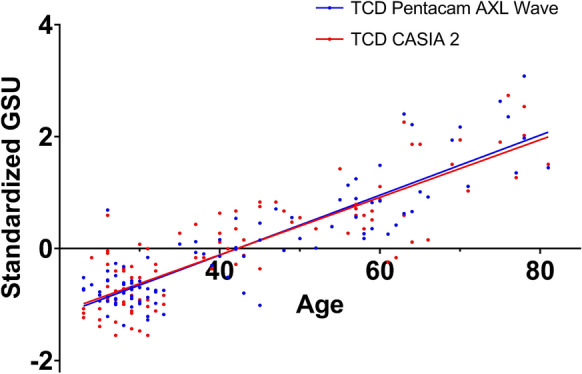
Results: The study included 96 healthy volunteers. The Casia 2 demonstrated high repeatability with ICC values exceeding 0.9 in all the corneal zones and lower CoV values compared to the Pentacam AXL Wave (ranging from 1.07% to 2.25% for Casia 2 and from 1.91% to 6.89% for Pentacam).95% LoA were within ± 2 standard deviation from the average mean except from zone 1 (± 2.42).However, the measurements showed a consistent bias among all the corneal zones. CD values were positively correlated with age, except for zone 1 with the Pentacam (p = 0.083).
Conclusions: The findings suggest that the Casia 2 can be a reliable tool for assessing corneal transparency in healthy individuals, however its measurements are not interchangeable with those provided by the Pentacam. The AS-OCT device may be more sensitive in detecting subtle age-related changes in CD within the central zone.
Keywords: AS-OCT; Corneal densitometry; Corneal opacity; Corneal transparency; Scheimpflug Imaging.
© 2024. The Author(s).
Conflict of interest statement
The authors declare no competing interests.
Figures
References
-
- Griffiths SN, Drasdo N, Barnes DA, Sabell AG (1986) Effect of epithelial and stromal edema on the light scattering properties of the cornea. Am J Optom Physiol Opt 63(11):888–894 - PubMed
-
- Braunstein RE, Jain S, McCally RL, Stark WJ, Connolly PJ, Azar DT (1996) Objective measurement of corneal light scattering after excimer laser keratectomy. Ophthalmology 103(3):439–443 - PubMed
-
- Krachmer JH (1978) Corneal endothelial dystrophy: a study of 64 families. Arch Ophthalmol 96(11):2036 - PubMed
-
- Smith GTH, Brown NAP, Shun-Shin GA (1990) Light scatter from the central human cornea. Eye 4(4):584–588 - PubMed
-
- Dohlman TH, Yin J, Dana R (2019) Methods for assessing corneal opacity. Semin Ophthalmol 34(4):205–210 - PubMed
Publication types
MeSH terms
LinkOut - more resources
Full Text Sources
Medical
Research Materials
Miscellaneous
