. 2024 Dec;115(12):3890-3901.
doi: 10.1111/cas.16344. Epub 2024 Sep 25.

Decreased PU.1 expression in mature B cells induces lymphomagenesis

Affiliations

Decreased PU.1 expression in mature B cells induces lymphomagenesis

Shinya Endo et al. Cancer Sci. 2024 Dec.

Abstract

Diffuse large B-cell lymphoma (DLBCL) is the most common subtype of lymphoma, accounting for 30% of non-Hodgkin lymphomas. Although comprehensive analysis of genetic abnormalities has led to the classification of lymphomas, the exact mechanism of lymphomagenesis remains elusive. The Ets family transcription factor, PU.1, encoded by Spi1, is essential for the development of myeloid and lymphoid cells. Our previous research illustrated the tumor suppressor function of PU.1 in classical Hodgkin lymphoma and myeloma cells. In the current study, we found that patients with DLBCL exhibited notably reduced PU.1 expression in their lymphoma cells, particularly in the non-germinal center B-cell-like (GCB) subtype. This observation suggests that downregulation of PU.1 may be implicated in DLBCL tumor growth. To further assess PU.1's role in mature B cells in vivo, we generated conditional Spi1 knockout mice using Cγ1-Cre mice. Remarkably, 13 of the 23 knockout mice (56%) showed splenomegaly, lymphadenopathy, or masses, with some having histologically confirmed B-cell lymphomas. In contrast, no wild-type mice developed B-cell lymphoma. In addition, RNA-seq analysis of lymphoma cells from Cγ1-Cre Spi1F/F mice showed high frequency of each monoclonal CDR3 sequence, indicating that these lymphoma cells were monoclonal tumor cells. When these B lymphoma cells were transplanted into immunodeficient recipient mice, all mice died within 3 weeks. Lentiviral-transduced Spi1 rescued 60% of the recipient mice, suggesting that PU.1 has a tumor suppressor function in vivo. Collectively, PU.1 is a tumor suppressor in mature B cells, and decreased PU.1 results in mature B-cell lymphoma development.

Keywords: DLBCL; PU.1; Spi1; diffuse large B‐cell lymphoma; lymphomagenesis.


Conflict of interest statement

The authors declare no conflict of interest. Koichi Akashi, Masao Matsuoka, and Yoshihiro Komohara are editorial board members of Cancer Science.

Figures

FIGURE 1
FIGURE 1
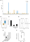
Spi1 is downregulated in human diffuse large B‐cell lymphoma (DLBCL) cells. (A) The Cancer Genome Atlas (TCGA) database showed that Spi1 was notably downregulated in various cancer types, specifically adrenocortical carcinoma (ACC), DLBCL, lung adenocarcinoma (LUAD), lung squamous cell carcinoma (LUSC), and thymoma (THYM) (* indicats P < 0.01). (B) In the TCGA database, Spi1 expression in DLBCL was significantly lower than that in normal tissue (* indicates P < 0.01). (C) The relative expression of Spi1 was measured in DLBCL cell lines, namely OCI‐Ly3 and SUDHL4. Peripheral blood mononuclear cells (PBMC) with CD19+ markers from a healthy individual served as a control for this measurement. (D) The expression of PU.1 was assessed on sorted CD19+ PBMC by flow cytometry. (E) Ratio of PU.1 expression in various cell lines to PBMCs using mean fluorescence intensity (MFI). (F) The GSE42203 database showed that ABC‐type DLBCL cell lines had reduced Spi1 expression compared to the GCB‐type cell lines.
FIGURE 2
FIGURE 2
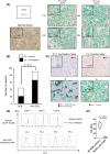
PU.1 is downregulated in human diffuse large B‐cell lymphoma (DLBCL) cells, particularly in non‐GCB type DLBCL cells. (A) The immunohistological analysis was done to study PU.1 in patients with DLBCL. Biopsy specimens were stained for CD19 and PU.1. Samples representing both high and low PU.1 expression in DLBCL, as well as a normal tonsil sample, are shown. (B) The data indicates a distinction in PU.1 expression levels between germinal center B‐cell‐like (GCB) and non‐GCB types of DLBCL. The non‐GCB DLBCL type showed a significantly decreased expression of PU.1 compared to GCB DLBCL (P < 0.01). (C) Double staining was done for PU.1 and Iba1. Interestingly, background macrophages had a strong expression of PU.1. (D) The downregulation of PU.1 in primary DLBCL cells was confirmed using flow cytometry. (E) The ratio of PU.1 expression in primary DLBCL cells compared to peripheral blood mononuclear cell (PBMC) CD19+. The findings showed a significant downregulation of PU.1 in non‐GCB type DLBCL cells (P < 0.0001).
FIGURE 3
FIGURE 3
Generation of conditional deletion of Spi1 in mature B cells via Cγ1‐Cre. (A) Schematic representation or design involving Spi1 F/F mice and Cγ1‐Cre mice (referred to as Cγ1‐Cre Spi1 F/F ). (B) The conditional knockout allele, presumably representing the knockout PU.1 gene, was detectable in specific tissues of the Cγ1‐Cre Spi1 F/F mice, namely the bone marrow and spleen. Interestingly, the specific primer set could only amplify this knockout allele, and this allele was only detectable in Cγ1‐Cre Spi1 F/F mice. (C) The survival rate of Cγ1‐Cre Spi1 F/F mice (n = 23) was comparable to that of the control Cγ1‐Cre mice (n = 26), suggesting no significant impact of the knockout on overall survival. (D) Various blood parameters such as white blood cell (WBC) counts, lymphocyte counts, hemoglobin (Hgb) levels, and platelet counts were similar between Cγ1‐Cre Spi1 F/F and control Cγ1‐Cre mice. (E) The spleen and liver of a sacrificed mouse. (F) Cγ1‐Cre Spi1 F/F mice exhibited infiltration of immature B lymphocytes in several organs, including the spleen, liver, lung, kidney, and salivary glands. Histological analyses such as hematoxylin–eosin (HE) staining, B220 staining, and Ki67 staining were conducted.
FIGURE 4
FIGURE 4
Conditional deletion of Spi1 in mature B cells induces diffuse large B‐cell lymphoma (DLBCL)‐like lymphoma that infiltrates into multiple organ tissues. (A) Incidence of tumors (represented in black) and splenomegaly (represented in white) in different mouse strains, observed in mice aged 18–24 months. (B) Representative tumors and splenomegaly. (C) Pathological findings of spleen on Spi1 F/F , Cγ1‐Cre Spi1 WT/WT , and Cγ1‐Cre Spi1 F/F mice. (D) hematoxylin–eosin (HE) staining and immunostaining with B220 and Ki67 of the intestinal tumor. (E) A dual immunostaining with B220 and PU.1 of spleen samples showed that the majority of B220+ cells (a marker for B lymphocytes) were found to be negative for PU.1 in Cγ1‐Cre Spi1 F/F mice.
FIGURE 5
FIGURE 5
Gene expression profiling revealed that diffuse large B‐cell lymphoma (DLBCL) cells in Cγ1‐Cre Spi1 F/F mice are monoclonal and have lymphoma‐related pathways. (A) BCR clonalities based on CDR3 sequences. Relative frequencies of CDR3 region detected in each sample are plotted. (B) A volcano plot of differentially expressed genes between B cells from Cγ1‐Cre Spi1 F/F mice and splenic B cells from Spi1 F/F mice. Fold change and adjusted P value (padj) are plotted for genes that are upregulated (yellow) or downregulated (blue) compared to the splenic B cells from Spi1 F/F mice. Representative genes related to malignant lymphoma are highlighted in red. (C) Over representation analysis (ORA) with differentially expressed genes in DLBCL cells from three Cγ1‐Cre Spi1 F/F mice compared with B cells in two control mice. Pathways related to malignant lymphoma are highlighted in red.
FIGURE 6
FIGURE 6
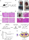
Lentivirally‐transduced Spi1 suppresses B lymphoma cell proliferation in recipient immunodeficient mice. (A) Schematic of the experimental strategy for transplantation of B lymphoma cells derived from Cγ1‐Cre Spi1 F/F mice to immunodeficient recipient mice (Rag2 −/− Jak3 −/− BALB/c). (B) After transplantation, the recipient mice exhibited symptoms of severe hepatosplenomegaly. (C) Pathological examinations of the spleen and liver of the immunodeficient recipient mice. (D) Immature lymphoma cells from P1 cells infiltrated into the spleen, central veins of the liver, and lungs in second serial‐transplanted recipient mouse. Hematoxylin and eosin staining of spleen, lung, and liver of the mice is shown. (E) Schematic of the experimental strategy for transplantation of B lymphoma cells derived from Cγ1‐Cre Spi1 F/F mice, which were transduced with a control lentivirus or Spi1‐expressing lentivirus. (F) Overall survival of recipient mice transplanted with vector only‐ or Spi1‐transduced lymphoma cells. (G) Graphical summary of this study.

References

    1. Sehn LH, Salles G. Diffuse large B‐cell lymphoma. N Engl J Med. 2021;384(9):842‐858. doi:10.1056/NEJMra2027612 - DOI - PMC - PubMed
    1. Coiffier B, Lepage E, Briere J, et al. CHOP chemotherapy plus rituximab compared with CHOP alone in elderly patients with diffuse large‐B‐cell lymphoma. N Engl J Med. 2002;346(4):235‐242. doi:10.1056/NEJMoa011795 - DOI - PubMed
    1. Pfreundschuh M, Trümper L, Osterborg A, et al. CHOP‐like chemotherapy plus rituximab versus CHOP‐like chemotherapy alone in young patients with good‐prognosis diffuse large‐B‐cell lymphoma: a randomised controlled trial by the MabThera International Trial (MInT) group. Lancet Oncol. 2006;7(5):379‐391. doi:10.1016/S1470-2045(06)70664-7 - DOI - PubMed
    1. Rosenwald A, Wright G, Chan WC, et al. The use of molecular profiling to predict survival after chemotherapy for diffuse large‐B‐cell lymphoma. N Engl J Med. 2002;346(25):1937‐1947. doi:10.1056/NEJMoa012914 - DOI - PubMed
    1. Meyer PN, Fu K, Greiner TC, et al. Immunohistochemical methods for predicting cell of origin and survival in patients with diffuse large B‐cell lymphoma treated with rituximab. J Clin Oncol. 2011;29(2):200‐207. doi:10.1200/JCO.2010.30.0368 - DOI - PMC - PubMed

MeSH terms