. 2024 Dec 2;30(23):5413-5429.
doi: 10.1158/1078-0432.CCR-24-1853.

Preclinical Evaluation of AZD6422, an Armored Chimeric Antigen Receptor T Cell Targeting CLDN18.2 in Gastric, Pancreatic, and Esophageal Cancers

Affiliations

Preclinical Evaluation of AZD6422, an Armored Chimeric Antigen Receptor T Cell Targeting CLDN18.2 in Gastric, Pancreatic, and Esophageal Cancers

Allison M Barrett et al. Clin Cancer Res. .

Abstract

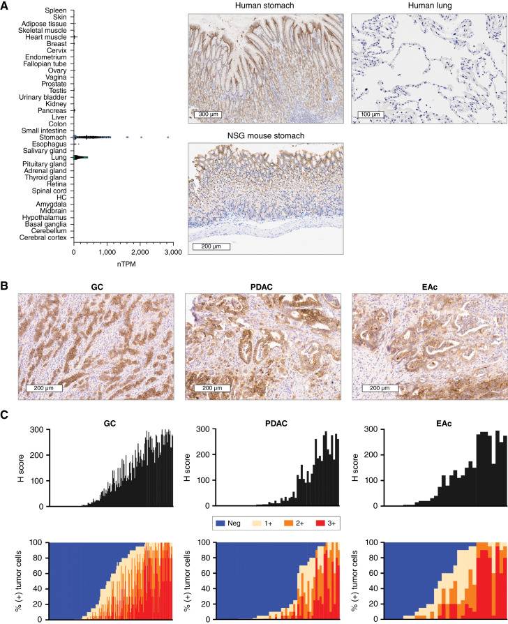
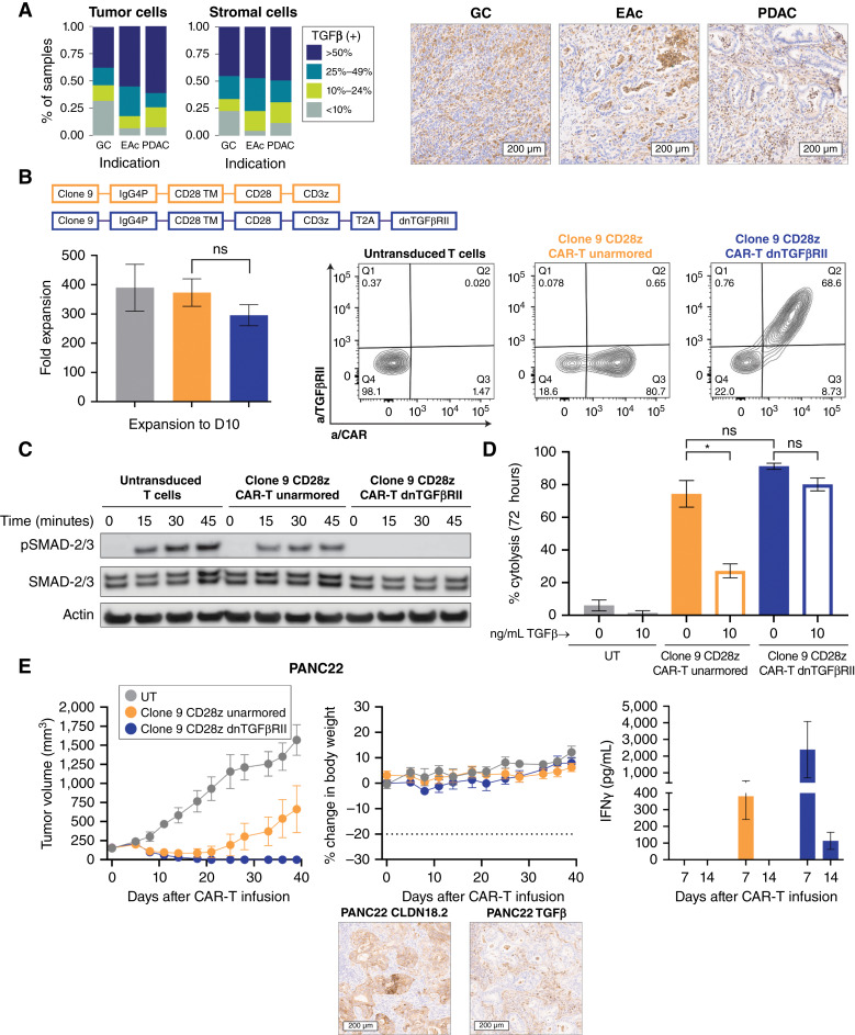
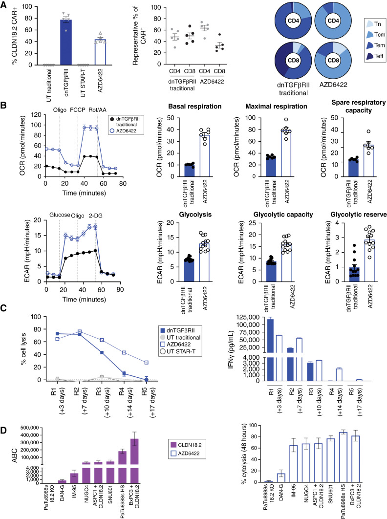
Purpose: Claudin 18.2 (CLDN18.2) is a surface membrane protein that is crucial for maintaining tight junctions in gastric mucosal cells and is highly expressed in gastric, esophageal, and pancreatic cancers. Thus, CLDN18.2 is suited for exploration as a clinical target for chimeric antigen receptor T-cell (CAR-T) therapy in these indications. Although CAR-T therapies show promise, a challenge faced in their development for solid tumors is the immunosuppressive tumor microenvironment, which is often characterized by the presence of immune and stromal cells secreting high levels of TGFβ. The addition of TGFβ armoring can potentially expand CAR-T activity in solid tumors. We report on the preclinical development of a CLDN18.2-targeting CAR-T therapy showing effectiveness in patient models with CLDN18.2-positive gastric, esophageal, and pancreatic tumors.

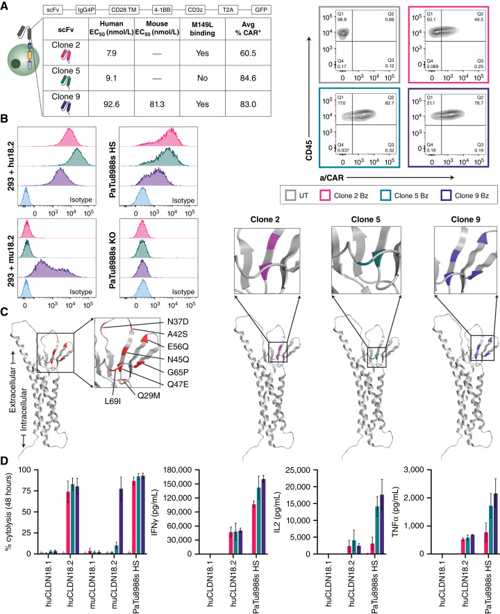
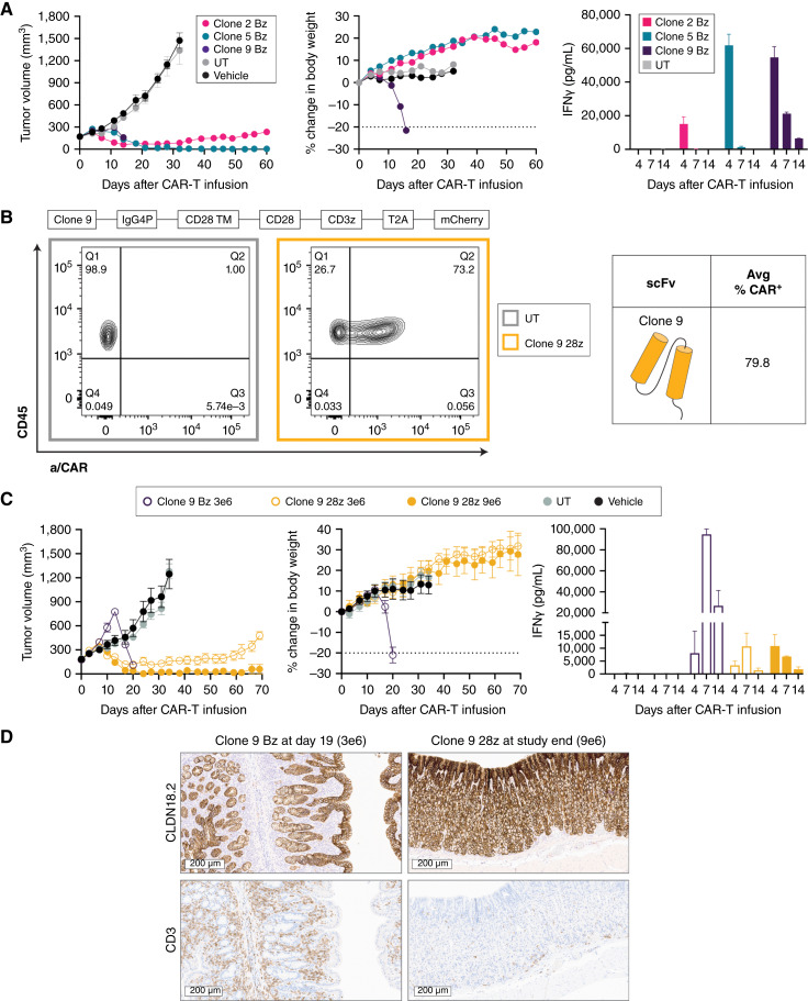
Experimental design: The lead lentivirus product contains a unique single-chain variable fragment; CD28 and CD3z costimulatory and signaling domains; and dominant-negative TGF-β receptor armoring, enhancing targeting and safety and counteracting suppression. We developed a shortened cell manufacturing process to enhance the potency of the final product AZD6422.

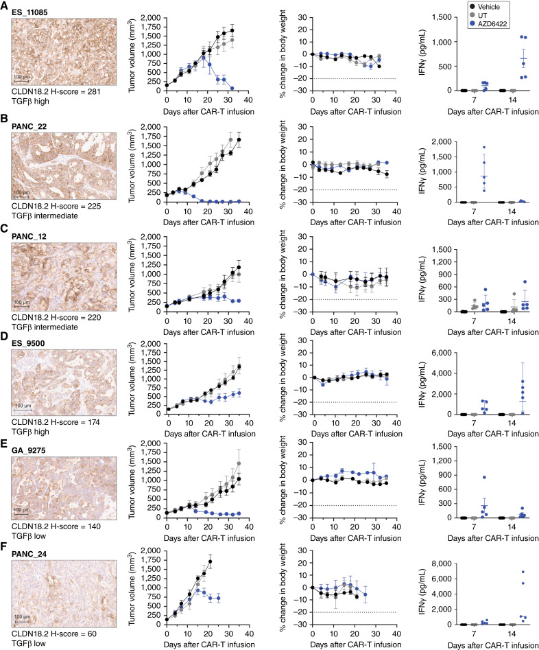
Results: AZD6422 exhibited significant antitumor activity and tolerability in multiple patient-derived tumor xenograft models with various CLDN18.2 and TGF-β levels, as determined by IHC. The efficacy of armored CAR-T cells in tumor models with elevated TGFβ was increased in vitro and in vivo. In vitro restimulation assays established greater persistence and cytolytic function of AZD6422 compared with a traditionally manufactured CAR-T.

Conclusions: AZD6422 was safe and efficacious in patient-derived, CLDN18.2-positive murine models of gastrointestinal cancers. Our data support further clinical development of AZD6422 for patients with these cancers.


Conflict of interest statement

All authors were employees of AstraZeneca when the work was completed and may have stock ownership, interests, and/or options in the company. A.M. Barrett reports a patent for US20240228610A9 pending. Z.T. Britton reports other support from AstraZeneca outside the submitted work; a patent for US20240228610A9 pending; and employment with AstraZeneca/MedImmune. R.A. Carrasco reports a patent for US20240228610A9 pending. S. Breen reports a patent for US20240228610A9 pending. A.L. Hatke reports a patent for US20240228610A9 pending. C. Yang reports a patent for US20240228610A9 pending. P.L. Martin reports other support from AstraZeneca during the conduct of the study; other support from AstraZeneca outside the submitted work; and employment with AstraZeneca and ownership of AZ stock. J.-M. Lapointe reports personal fees from AstraZeneca during the conduct of the study. M. Cobbold reports other support from AstraZeneca during the conduct of the study. G. Moody reports a patent for US20240228610A9 pending to AstraZeneca, as well as employment with AstraZeneca LP and ownership of AstraZeneca stock. E.E. Bosco reports other support from AstraZeneca during the conduct of the study. No disclosures were reported by the other authors.

Figures

Figure 1.
Figure 1.
Expression and prevalence of CLDN18.2 in normal tissues and GI adenocarcinomas. A, Normal tissue RNA expression of CLDN18 [represented as normalized transcripts per million (TPM); Human Protein Atlas, GTEx dataset]. HC, hippocampal formation. CLDN18.2 expression on representative images of the human stomach, NSG mouse stomach, and human lung are shown. B, CLDN18.2 expression on representative samples of gastric cancer (H-score = 249), PDAC (H-score = 175), and esophageal adenocarcinoma (H-score = 175). C, Prevalence and intensity of CLDN18.2 expression on tumor samples derived from patients with gastric cancer (n = 185), PDAC (n = 61), and esophageal adenocarcinoma (n = 32). Data are shown as H-scores and as the percentages of tumor cells at each level of intensity (1+, 2+, and 3+). EAc, esophageal adenocarcinoma; GC, gastric cancer.
Figure 2.
Figure 2.
Development and evaluation of CLDN18.2-targeting CAR-T cells in vitro. A, Schematic representation of second-generation CAR-T lentivirus design, which includes a 4-1BB costimulatory domain (Bz). The table shows for each CLDN18.2-reactive clone the relative binding affinity (human and mouse), reactivity to mutant CLDN18.2 (M149L), and average transduction efficiency (CAR+, day 9) of multiple healthy donors. Representative FC plots of CAR surface expression at day 9 after lentivirus transduction were compared with UT control for a single donor. B, CLDN18.2 cell surface expression of various cell lines as determined by FC with 5 μg/mL CLDN18.2-reactive clones compared with nonspecific isotype antibody (R347). C, Epitope characterization of CLDN18.2-reactive clones. The AlphaFold structure on the far left (red) represents all sites of point mutation in HEK293 cells that vary between CLDN18.1 and CLDN18.2 in the first extracellular loop; the color-coded diagrams represent sites that influence respective clone binding. D, Percent cytolysis of HEK293 + huCLDN18.1, HEK293 + huCLDN18.2, HEK293 + muCLDN18.1, HEK293 + muCLDN18.2, and PaTu8988s HS cells determined by xCELLigence RTCA assay after 48 hours of co-culture with CLDN18.2 CAR-T cells at a 1:1 E:T ratio. The supernatants from the xCELLigence assay were collected at 24 hours for cytokine assessment (Meso Scale Discovery) assay. All data represent mean ± SEM of replicate experiments.
Figure 3.
Figure 3.
Evaluation of CLDN18.2-targeting CAR-T cells in vivo. A, NSG mice bearing PaTu8988s HS xenografts (CLDN18.2 H-score = 268) were dosed by tail vein with 9e6 CAR+ CLDN18.2 Bz CAR-T cells; total T-cell infusion number was matched across groups. Tumor volume and body weight were measured biweekly (n = 9). Serum levels of IFNγ were measured at 4, 7, and 14 days after infusion (n = 3). B, Schematic representation of a second-generation CAR-T design modified to replace 4-1BB with a CD28 costimulatory domain (28z). The average transduction efficiency (CAR+, day 9) of multiple healthy donors for clone 9 28z is shown. Representative FC plots of CAR surface expression at day 9 after lentivirus transduction were compared with UT control for a single donor. C, NSG mice bearing PaTu8988s HS xenografts were dosed as described in A with clone 9 CD28z or Bz CAR-T (n = 6) at indicated doses. Serum levels of IFNγ were measured at 4, 7, and 14 days after infusion (n = 3). D, Representative images of CLDN18.2 (top row) and CD3 (bottom row) staining in the stomachs of mice dosed with clone 9 CAR-T cells from C at indicated time points. All data represent mean ± SEM of replicate experiments or animals.
Figure 4.
Figure 4.
Rationale for selection of TGFβ armoring and proof of mechanism in vitro and in vivo. A, Quantitative analysis of intensity and prevalence and representative images of TGFβ staining in a subset of patient tumor samples [gastric cancer (GC), n = 130; esophageal adenocarcinoma (EAc), n = 15; PDAC, n = 71]. Data represent the pooled scores of tumor and stromal compartments. B, Schematic representation of second-generation CAR-T lentivirus design, including an IgG4P hinge, CD28 transmembrane, CD28 costimulatory domain, and CD3z (unarmored CAR-T cells) or additional T2A self-cleaving peptide and dnTGFβRII (armored CAR-T cells). Average fold expansion across multiple healthy donors is shown. There was no significant difference in expansion across groups (one-way ANOVA). Representative FC plots show CAR and TGFβRII surface expression at day 10 after lentivirus transduction compared with UT control. C, FACS-purified, serum-starved CAR-T cells were stimulated with 1 ng/mL rhTGFβ for various time periods. Western blotting was used to determine protein levels of p-SMAD2/3 and total SMAD2/3; β-actin was used as the loading control. D, Percent cytolysis of BxPC3 + CLDN18.2 cells as determined by xCELLigence RTCA assay after 72 hours of co-culture at a 1:1 ratio with CLDN18.2 CAR-T cells in the presence or absence of 10 ng/mL rhTGFβ. Results were analyzed by using paired t tests. E, NSG mice bearing pancreatic PDX (PANC22, H-score = 225, TGFβ intermediate) were dosed by tail vein with 3e6 CAR+ unarmored or dnTGFβRII CLDN18.2 CAR-T cells; the total T-cell infusion number was matched across groups. Tumor volume and body weight were measured biweekly (n = 5). Serum levels of IFNγ were measured at 7 and 14 days after infusion (n = 3). Representative images of CLDN18.2 and TGFβ staining IHC expression (20× scan) are shown. All data represent mean ± SEM of replicate experiments or animals.
Figure 5.
Figure 5.
Optimized manufacturing protocol, STAR-T, for the generation of the CAR-T product. A, Baseline characteristics of donor-matched dnTGFβRII CAR-T cells with traditional manufacture (day 10) vs. AZD6422 (day 4), including CAR+ expression, percent CD4 and CD8 expression, and T-cell phenotypic status as determined by cell surface expression of CCR7 and CD45RO. Results are shown for naïve (CCR7+/CD45RO, Tn), central memory (CCR7+/CD45RO+, Tcm), effector memory (CCR7/CD45RO+, Tem), and effector (CCR7/CD45RO, Teff) cells. Data are shown as mean ± SEM of representative donors. B, Comparison of bioenergetic profiles of traditionally manufactured dnTGFβRII CAR-T cells vs. AZD6422. Spare respiratory capacity was determined as the differential between basal and maximum respiration. 2-DG, 2-deoxy-D-glucose; ECAR, extracellular acidification rate; FCCP, carbonyl cyanide p-trifluoro-methoxyphenyl hydrazone; OCR, oxygen consumption rate; Oligo, oligomycin; Rot/AA, rotenone and antimycin A. C, Serial restimulation assay to examine cytotoxicity and persistence of dnTGFβRII CAR-T cells and AZD6422. CAR-T cells were co-cultured at a ratio of 1:2 with BXPC3 + CLDN18.2, tumor lysis was measured every 3 to 4 days, and IFNγ was profiled at 24 hours after each new co-culture. Representative of multiple donors. D, Results of quantitative FC to determine cell surface expression of CLDN18.2 across multiple cancer cell lines. Percent cytolysis was determined by xCELLigence RTCA assay after 48 hours of co-culture with AZD6422 at an E:T ratio of 1:1. Data represent mean ± SEM of replicate experiments.
Figure 6.
Figure 6.
In vivo antitumor activity of AZD6422 in PDX models of gastric cancer, PDAC, and esophageal adenocarcinoma. Activity and tolerability of AZD6422 are shown in various PDX models of esophageal adenocarcinoma (A, ES11085; D, ES_9500), gastric cancer (E, GA_9275), and PDAC (B, PANC_22; C, PANC_12; F, PANC_24). Each model was selected to represent a range of CLDN18.2 (shown at 10× scan) and TGFβ expression. NSG MHC-DKO mice received a single tail-vein infusion of 1e6 AZD6422, donor-matched UT, or vehicle when the average tumor volume reached 150 mm3. Tumor volumes and body weights were measured biweekly until study completion on day 35, and blood was collected for cytokine analysis on days 7 and 14. Data are shown as mean ± SEM (n = 5).

References

    1. Kayali S, Marabotto E, Giannini E. Gastrointestinal tract cancers, an increasing burden of the modern era: epidemiology and prevention. Cancers (Basel) 2023;15:4634. - PMC - PubMed
    1. Sung H, Ferlay J, Siegel RL, Laversanne M, Soerjomataram I, Jemal A, et al. . Global cancer statistics 2020: GLOBOCAN estimates of incidence and mortality worldwide for 36 cancers in 185 countries. CA Cancer J Clin 2021;71:209–49. - PubMed
    1. Guan WL, He Y, Xu RH. Gastric cancer treatment: recent progress and future perspectives. J Hematol Oncol 2023;16:57. - PMC - PubMed
    1. Mou P, Ge Q-H, Sheng R, Zhu T-F, Liu Y, Ding K. Research progress on the immune microenvironment and immunotherapy in gastric cancer. Front Immunol 2023;14:1291117. - PMC - PubMed
    1. Luo W, Wang J, Chen H, Qiu J, Wang R, Liu Y, et al. . Novel strategies optimize immunotherapy by improving the cytotoxic function of T cells for pancreatic cancer treatment. Cancer Lett 2023;576:216423. - PubMed

MeSH terms

Grants and funding